Using TTchem-seq for profiling nascent transcription and measuring transcript elongation
https://doi.org/10.1038/s41596-019-0262-3
https://doi.org/10.1038/s41596-019-0262-3
TT for nascent Using chem-seq profiling transcription and measuring transcript elongation
Lea H. Gregersen1, Richard Mitter2 and Jesper Q. Svejstrup1*
The dynamics of transcription can be studied genome wide by high-throughput sequencing of nascent and newly synthesized RNA. 4-thiouridine (4SU) labeling in vivo enables the specific capture of such new transcripts, with 4SU residues being tagged by biotin linkers and captured using streptavidin beads before library production and highthroughput sequencing. To achieve high-resolution profiles of transcribed regions, an RNA fragmentation step before biotin tagging was introduced, in an approach known as transient transcriptome sequencing (TT-seq). We recently introduced a chemical approach for RNA fragmentation that we refer to as TTchem-seq. We describe how TTchem-seq can be used in combination with transient inhibition of early elongation using the reversible CDK9 inhibitor, 5,6dichlorobenzimidazole 1-β-D-ribofuranoside (DRB), to measure RNA polymerase II (RNAPII) elongation rates in vivo, a technique we call DRB/TTchem-seq. Here, we provide detailed protocols for carrying out TTchem-seq and DRB/TTchem-seq, including computational analysis. Experiments and data analysis can be performed over a period of 10–13 d and require molecular biology and bioinformatics skills.
使用化学测序(chem-seq)分析转录和测量转录本延伸的TT方法
Lea H. Gregersen1, Richard Mitter2 and Jesper Q. Svejstrup1*
Steady-state RNA levels are influenced by transcription rate, co-transcriptional processing, RNA modification and turnover. Measurement of steady-state levels is often insufficient to study the dynamic transcriptional response to stress or stimuli. Metabolic labeling of cells with 4-thiouridine (4SU) combined with high-throughput sequencing provides a convenient method to capture nascent (RNA polymerase–associated) and newly synthesized RNA transcripts in vivo. A short 5–15 min incubation with 4SU selectively labels newly transcribed RNA1,2. The ease of 4SU labeling, combined with its high reproducibility, has made it a popular technique for studying transcription dynamics and has led to the development of several methodologies1–10, the most recent of which is transient transcriptome sequencing (TT-seq), first described by the Cramer laboratory9. Here, we describe a detailed protocol for TTchem-seq that enables obtaining high-resolution transcriptome profiles of nascent and newly transcribed RNA by using hydrolysis rather than sonication to fragment RNA. We also provide a protocol for inferring RNAPII elongation rates in vivo, by combining DRB-mediated RNAPII inhibition with TTchem-seq11, in what we term DRB/TTchem-seq. The experimental parts of the protocol require basic knowledge of molecular biology and tissue culture. The computational analysis requires prior knowledge of data analysis, as well as the R and Bash programming languages.
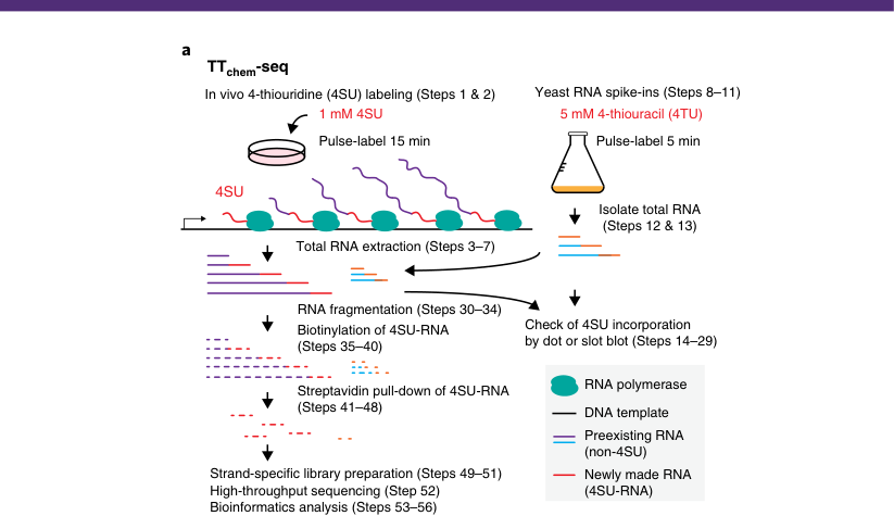
An overview of the TTchem-seq and DRB/TTchem-seq protocols is shown in Fig. 1. In both cases, cells are pulse-labeled with 4SU in vivo, and total RNA is extracted. In parallel, yeast cells are labeled with 4-thiouracil (4TU); a small amount of this labeled yeast RNA is spiked into the mammalian RNA to serve as a normalization control. Next, the RNA is fragmented by controlled base hydrolysis. Only RNA regions transcribed within the short pulse will contain 4SU residues, which are selectively captured after RNA fragmentation. This ensures that only transcript regions that have recently been produced are mapped. By contrast, protocols without the RNA fragmentation step capture any RNA transcript containing 4SU residues at any given position within the transcript. The pool of
1Mechanisms of Transcription Laboratory, The Francis Crick Institute, London, UK. 2Bioinformatics and Biostatistics, The Francis Crick Institute, London, UK. *e-mail: jesper.svejstrup@crick.ac.uk
a, TTchem-seq: In vivo 4-thiouridine (4SU) labeling (Steps 1 & 2), Yeast RNA spike-ins (Steps 8–11): 1 mM 4SU, 5 mM 4-thiouracil (4TU), Pulse-label 15 min, Pulse-label 5 min, 4SU: Isolate total RNA (Steps 12 & 13), Total RNA extraction (Steps 3–7), RNA fragmentation (Steps 30–34), Biotinylation of 4SU-RNA Check of 4SU incorporation (Steps 35–40) by dot or slot blot (Steps 14–29), Streptavidin pull-down of 4SU-RNA, RNA polymerase DNA template Preexisting RNA (non-4SU), Strand-specific library preparation (Steps 49–51), Newly made RNA, High-throughput sequencing (Step 52) (4SU-RNA), Bioinformatics analysis (Steps 53–56).
b DRB/TTchem-seq DRB incubation for 3.5 h RNAPII synchronized close to the TSS
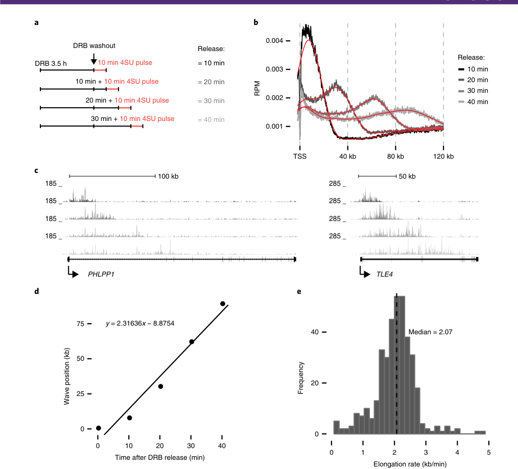
10 min release, 20 min release, 30 min release, 40 min release: (10 min 4SU), (10 min+10 min 4SU), (20 min+10 min 4SU), (30 min+10 min 4SU).
Fig. 1 | Overview of TTchem-seq and DRB/TTchem-seq. a, Detailed overview of the workflow for TTchem-seq, including generation of yeast spike-in normalization controls. Nascent RNA is labeled in vivo by addition of 4SU directly to the tissue culture medium. The reaction is stopped by TRIzol, and total RNA is extracted and fragmented by controlled base hydrolysis. 4SU residues in the fragmented RNA are biotinylated and used for streptavidin pulldown of 4SU-containing RNA. b, Principle of DRB/TTchem-seq. Early RNAPII elongation is inhibited by DRB, which can be removed by PBS washes and medium replacement. Time-dependent release of RNAPII after DRB treatment, coupled with TTchem-seq, will label newly synthesized RNA as the RNAPII wave peak progresses throughout the gene body.
fragmented mammalian and yeast RNA containing 4SU is then biotinylated using a biotin linker that reacts specifically with 4SU residues. This enables a high-stringency streptavidin purification step to separate newly transcribed 4SU-labeled RNA from preexisting non-labeled RNA, before strand-specific library preparation for high-throughput sequencing. Because there is no selection for polyadenylated transcripts, TTchem-seq captures regions of protein-coding and non-coding transcripts equally. Thus, TTchem-seq is an excellent method for obtaining high-resolution transcriptome profiles across protein-coding genes and long non-coding RNAs (lncRNAs), as well as for capturing short-lived RNA intermediates such as antisense transcripts and transcript regions downstream of the polyadenylation sites. TTchem-seq can furthermore be adapted to measure RNAPII elongation rates by taking advantage of inhibitor-mediated synchronization of RNAPII molecules close to the transcription start site, followed by release from inhibition as described in our DRB/TTchem-seq protocol (Fig. 1b).
1Mechanisms of Transcription Laboratory, The Francis Crick Institute, London, UK. 2Bioinformatics and Biostatistics, The Francis Crick Institute, London, UK. *e-mail: jesper.svejstrup@crick.ac.uk
a, TTchem-seq: 体内4-硫代尿苷 (4SU) 标记(步骤1和2),酵母RNA加入对照(步骤8–11):1 mM 4SU,5 mM 4-硫脲 (4TU),脉冲标记 15 min,脉冲标记 5 min,4SU:分离总RNA(步骤12和13),总RNA提取(步骤3–7),RNA片段化(步骤30–34),4SU-RNA的生物素化,通过点或槽印检测4SU掺入情况(步骤35–40)(步骤14–29),4SU-RNA的链霉亲和素下拉法,RNA聚合酶DNA模板,预先存在的RNA(非4SU),特异性文库制备(步骤49–51),新合成的RNA,高通量测序(步骤52)(4SU-RNA),生物信息学分析(步骤53–56)。
b DRB/TTchem-seq 在TSS附近同步的RNAPII进行DRB孵育 3.5 h
10 min释放,20 min释放,30 min释放,40 min释放:(10 min 4SU),(10 min+10 min 4SU),(20 min+10 min 4SU),(30 min+10 min 4SU)。
fragmented mammalian and yeast RNA containing 4SU is then biotinylated using a biotin linker that reacts specifically with 4SU residues. This enables a high-stringency streptavidin purification step to separate newly transcribed 4SU-labeled RNA from preexisting non-labeled RNA, before strand-specific library preparation for high-throughput sequencing. Because there is no selection for polyadenylated transcripts, TTchem-seq captures regions of protein-coding and non-coding transcripts equally. Thus, TTchem-seq is an excellent method for obtaining high-resolution transcriptome profiles across protein-coding genes and long non-coding RNAs (lncRNAs), as well as for capturing short-lived RNA intermediates such as antisense transcripts and transcript regions downstream of the polyadenylation sites. TTchem-seq can furthermore be adapted to measure RNAPII elongation rates by taking advantage of inhibitor-mediated synchronization of RNAPII molecules close to the transcription start site, followed by release from inhibition as described in our DRB/TTchem-seq protocol (Fig. 1b).
Development of TTchem-seq
In recent years, several modifications and technical improvements have been introduced to protocols utilizing 4SU labeling4,7,9,12–14. The protocol described here details the latest developments aimed at obtaining high-resolution transcriptome profiles of nascent and newly synthesized RNA. Critical alterations to the original 4SU protocols are the addition of an RNA fragmentation step before biotinylation and pull-down of the labeled RNA. By including this fragmentation step, only newly produced RNA regions are isolated, enabling the experimenter to pinpoint precisely where transcription is taking place within a transcriptional unit. A similar approach was developed by the Cramer lab, but that method, termed TT-seq, uses sonication to fragment the RNA9. Because our protocol uses a different fragmentation method from that of Cramer and colleagues, we refer to it as TTchem-seq. We find that controlled base hydrolysis of the RNA results in a narrow size distribution of RNA fragments and that fragment length can easily be adjusted by simply increasing or decreasing the time of the base hydrolysis. Nascent RNA makes up a very small proportion of the total RNA within a cell, which mainly consists of stable ribosomal RNA (rRNA). Depending on labeling times, the 4SU-containing RNA will range from 0.2 to 0.7% of the total RNA and will be primarily generated by RNAPII. By contrast, the vast majority of total RNA consists of rRNA, owing to the longer half-life of rRNA relative to mRNA. A highly convenient feature of 4SU is that it has increased reactivity toward activated disulfides as compared to other nucleotides4,15. Thus, RNA containing 4SU residues can be captured by the addition of biotin linkers containing such activated disulfides, which will react specifically with the 4SU thiol group and enable enrichment of 4SU-containing RNA by streptavidin beads. Original protocols for capturing 4SU-labeled RNA used EZ-Link HPDP-Biotin to link a biotin tag to 4SU residues3,6,15. However, a MTSEA BIOTIN-XX linker that offers increased reactivity with shorter reaction times was recently described4. We have successfully used both types of biotin linkers and found that MTSEA BIOTIN-XX indeed results in a greater capture of 4SU-containing RNA (data not shown). The following protocol will therefore detail the use of this linker only. For reaction conditions using the EZ-Link HPDP-Biotin linker, refer to previously published protocols3,6.
Adaptation of TTchem-seq to study RNAPII elongation rates: DRB/TTchem-seq
One particularly useful adaptation of TTchem-seq is its use to measure RNAPII elongation rates when combined with DRB treatment (Fig. 1b). DRB has previously been used to measure genome-wide RNAPII transcript elongation rates in vivo by us11,16 and the Oren lab5,17. DRB inhibits the kinase activity of CDK9, which is part of the P-TEFb complex and is required for phosphorylation of Spt5, as well as the RNAPII C-terminal domain18,19. Lack of CDK9 activity results in a failure of newly initiated RNAPII to progress to the elongation phase, while permitting mature elongation complexes to complete transcription. DRB thus, in effect, synchronizes the transcription cycle by reversibly blocking new transcript elongation. In combination with TTchem-seq, the progression of RNAPII into the gene body can then be tracked in a time-resolved manner upon DRB release to determine the speed of RNAPII elongation rates in DRB/TTchem-seq (Fig. 1b)11.
将 TTchem-seq 应用于研究 RNAPII 延伸速率:DRB/TTchem-seq
One particularly useful adaptation of TTchem-seq is its use to measure RNAPII elongation rates when combined with DRB treatment (Fig. 1b). DRB has previously been used to measure genome-wide RNAPII transcript elongation rates in vivo by us11,16 and the Oren lab5,17. DRB inhibits the kinase activity of CDK9, which is part of the P-TEFb complex and is required for phosphorylation of Spt5, as well as the RNAPII C-terminal domain18,19. Lack of CDK9 activity results in a failure of newly initiated RNAPII to progress to the elongation phase, while permitting mature elongation complexes to complete transcription. DRB thus, in effect, synchronizes the transcription cycle by reversibly blocking new transcript elongation. In combination with TTchem-seq, the progression of RNAPII into the gene body can then be tracked in a time-resolved manner upon DRB release to determine the speed of RNAPII elongation rates in DRB/TTchem-seq (Fig. 1b)11.
Comparison with other methods
Traditionally, transcription has been studied using RNAPII chromatin immunoprecipitation (ChIP); more recently, nucleotide-resolution native elongating transcript sequencing (NET-seq) has been used20–22. ChIP measures RNAPII occupancy and location on the basis of isolation of chromatin, typically followed by nuclease digestion and enrichment of transcribed regions using antibodies against either total or phosphorylated forms of RNAPII. Similarly, NET-seq and mNET-seq involve isolation of chromatin and, in the case of mNET-seq, an immunoprecipitation step using antibodies against RNAPII20–22. However, in both cases RNA rather than the DNA associated with RNAPII is isolated and used to infer RNAPII occupancy. In yeast, photoactivatable ribonucleoside-enhanced crosslinking and immunoprecipitation (PAR-CLIP) or modification crosslinking and analysis of cDNA (mCRAC) have also been used to map RNAPII binding to RNA genome wide23,24. Both PARCLIP and mCRAC capture RNA associated with RNAPII at a nucleotide resolution; however, unlike NET-seq, both methods include a UV-induced RNA–protein crosslinking step before RNAPII immunoprecipitation. As an alternative approach, transcription activity can be studied using techniques to directly label and isolate nascent RNA and newly transcribed RNA. One such method is global run-on and sequencing (GRO-seq), which relies on isolation of nuclei and a ‘run-on’ transcription reaction25. However, the isolation of nuclei is a relatively time-consuming procedure and
may introduce bias with short-scale time point measurements (a 4SU-labeling pulse in the range of 5–15 min), especially if many samples need to be processed in parallel. Moreover, the transcription reaction performed in GRO-seq is, in effect, an in vitro reaction, involving addition of nucleotides to isolated nuclei to prompt RNA polymerases to label preexisting transcripts. One major advantage of TTchem-seq and TT-seq for studying nascent transcription is that all transcript labeling is carried out in vivo. This eliminates the need to isolate nuclei and minimizes any variability or cellular stress that might be introduced during the transcription reaction. In addition, the short 4SU-labeling reaction is stopped by the addition of TRIzol directly to mammalian cells, providing a fast and easy way to control the exact duration of labeling. This is particularly important when multiple samples need to be directly compared. Another notable difference is that the run-on buffer used in GRO-seq usually contains sarkosyl, which can release promoter-paused RNAPII and may remove regulatory factors bound to the polymerase26. Similarly, ChIP, NET-seq, PAR-CLIP and mCRAC also capture RNA associated with paused or inactive RNAPII molecules in chromatin. By contrast, TTchem-seq and TT-seq capture only nascent or newly transcribed RNA from actively elongating RNAPII complexes because the labeling is performed in vivo without perturbation of the transcription process.
As an alternative to biotin tagging and streptavidin-mediated enrichment of 4SU-containing RNA, 4SU residues can be chemically converted into cytidine analogs, which results in nucleotide conversion in the sequencing reads7,27,28. This enables direct detection of 4SU residues from total RNAseq by identification of T>C transitions in the sequencing reads. However, because nascent transcripts make up a very small fraction of total RNA, only an exceedingly small percentage of transcripts will contain 4SU residues (and thus cytidine after conversion) after a 5–15 min labeling. Because these approaches lack an enrichment step for 4SU-containing RNA, they require much greater sequencing read depth to obtain sufficient coverage of 4SU/cytidine residues and this substantially increases the costs associated with sequencing. To our knowledge, these approaches have been used only with longer labeling times (45 min as the shortest labeling time7), which makes them less suited to study nascent transcription.
The idea of using CDK9 inhibitors to study the dynamics of RNAPII elongation in vivo is not new. Non-reversible CDK9 inhibitors such as DRB, triptolide and flavopiridol have been used to measure the time-dependent movement of RNAPII elongation complexes in gene bodies5,16,29–31. Most of the initial approaches involved the isolation of nuclei and in vitro run-on reactions; however, the Oren lab combined DRB treatment with non-fragmented 4SU-seq (termed 4sUDRB-seq) to measure RNAPII progression 4 and 8 min after DRB release5,17. The DRB/TTchem-seq protocol outlined here offers several advantages compared to these pioneering approaches. First, the RNA fragmentation step results in well-defined wave peaks of RNAPII elongation complexes (regions actively transcribed by the wave of RNAPII complexes released from DRB inhibition; see also Fig. 1b), which can be used to computationally track RNAPII progression, compared to the ‘boundary detection’ used by Fuchs et al.5,17, which is more sensitive to background signals downstream of the RNAPII wave. In addition, we measure nascent transcription at four time points after DRB release. This enables us to take information from multiple time points into account by fitting a linear regression to calculate the elongation rate. We also adapt the use of the MTSEA BIOTIN-XX linker, resulting in increased sensitivity, which is particularly important when using the short-duration 4SU labeling required for this approach. Finally, we use yeast RNA spike-in for global normalization and to control for equal biotin tagging, 4SU pull-down and library preparation between samples (as discussed below). We use a fitted spline approach to identify the RNAPII wave peak. Other methods, for example, those using GRO-seq, have either used hidden Markov models (HMMs) or simply identified stretches where read coverage dropped as compared to that of upstream regions16,32. Previously reported global elongation rates obtained using DRB/GRO-seq were in the range of 2–4 kb/min16, whereas elongation rates calculated by 4sUDRB-seq ranged from 2–6 kb/min5. On the basis of DRB/TTchem-seq, we find that most genes have an elongation rate ~2 kb/min (Anticipated results), although we do observe some variation in elongation rates between genes.
Comparison with other methods
Traditionally, transcription has been studied using RNAPII chromatin immunoprecipitation (ChIP); more recently, nucleotide-resolution native elongating transcript sequencing (NET-seq) has been used20–22. ChIP measures RNAPII occupancy and location on the basis of isolation of chromatin, typically followed by nuclease digestion and enrichment of transcribed regions using antibodies against either total or phosphorylated forms of RNAPII. Similarly, NET-seq and mNET-seq involve isolation of chromatin and, in the case of mNET-seq, an immunoprecipitation step using antibodies against RNAPII20–22. However, in both cases RNA rather than the DNA associated with RNAPII is isolated and used to infer RNAPII occupancy. In yeast, photoactivatable ribonucleoside-enhanced crosslinking and immunoprecipitation (PAR-CLIP) or modification crosslinking and analysis of cDNA (mCRAC) have also been used to map RNAPII binding to RNA genome wide23,24. Both PARCLIP and mCRAC capture RNA associated with RNAPII at a nucleotide resolution; however, unlike NET-seq, both methods include a UV-induced RNA–protein crosslinking step before RNAPII immunoprecipitation. As an alternative approach, transcription activity can be studied using techniques to directly label and isolate nascent RNA and newly transcribed RNA. One such method is global run-on and sequencing (GRO-seq), which relies on isolation of nuclei and a ‘run-on’ transcription reaction25. However, the isolation of nuclei is a relatively time-consuming procedure and
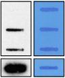
The success of TTchem-seq is dependent on efficient cellular uptake and labeling of newly synthesized RNA with 4SU. It is therefore critical to check the efficiency of RNA 4SU incorporation before the streptavidin pull-down and library preparation by performing a dot or slot blot assay (Fig. 2a).
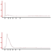
The size distribution of fragmented RNA can be checked by Bioanalyzer, either before or after the streptavidin pull-down (Fig. 2b). Alternatively, a denaturing agarose gel can be used to determine the size range of RNA fragments before the pull-down (Supplementary Fig. 2a). We aim for an RNA size distribution between 25 and 500 nt for both TTchem-seq and DRB/TTchem-seq. The size distribution of RNA fragments can easily be controlled by simply increasing or decreasing the time the RNA is treated with sodium hydroxide (Supplementary Fig. 2a). It is important to keep the size range of the RNA fragments in mind for subsequent steps in the protocol. For instance, many column-based RNA purification kits select for fragments >200 nt. To avoid loss of fragments <200 nt, it is necessary to increase the ethanol amount when using the Qiagen minElute columns to clean up 4SU-RNA after streptavidin purification (Supplementary Fig. 2b).
Biotinylation and streptavidin pull-down of 4SU-RNA
The use of methanethiosulfonate (MTS)-biotin to tag 4SU-RNA offers increased reactivity toward thiols, resulting in a >95% conversion rate of 4SU residues to biotin-4SU, as compared with <20% for HPDP-Biotin4 . We find that μMACS streptavidin beads, in combination with μColumns and a high-salt wash to rigorously enrich for 4SU-RNA, results in exceedingly low amounts of crosscontamination from non-labeled RNA. Using the conditions detailed in the protocol below, we purify <1% of RNA from a non-4SU-labeled background sample as compared with cells treated for 15 min with 1 mM 4SU (Fig. 2b). The RNA fragmentation step is important to achieving this because it has been reported to decrease background levels when using MTS-biotin13 . It is critical to confirm the efficiency of 4SU-RNA enrichment and the size of the RNA fragments before library preparation by Bioanalyzer (Fig. 2b).
Although it is possible to perform rRNA depletion before library preparation, this is not strictly necessary, because most transcripts synthesized within the 5- to 15-min pulse actually originate from RNAPII-transcribed transcripts. For both TTchem-seq and DRB/TTchem-seq libraries, we thus consistently observe <0.2% of reads mapping to rRNA. The sequencing depth required for TTchemseq libraries is generally higher than that for mRNA-seq libraries, owing to the higher sequence complexity caused by the high proportion of non-coding regions (including introns) included in TTchem-seq. Typically, we sequence each library to a depth of ~50–70 million reads. Single-end sequencing is sufficient to measure newly synthesized transcripts using TTchem-seq and RNAPII elongation rates using DRB/TTchem-seq. However, paired-end sequencing can be used to gain information about co-transcriptional splicing occurring within the time frame of the 4SU pulse.
The most important considerations and limitations of TTchem-seq and DRB/TTchem-seq are as follows:
Because RNA fragmentation results in fragments ranging from 25 to 500 nt, this is the resolution at which regions of active transcription can be detected. If higher resolution is desired, the time of base hydrolysis can be increased to obtain smaller fragments (Supplementary Fig. 2a). This could, for instance, be required to uniquely assign reads to closely spaced transcription units. Beyond the size-exclusion columns used to clean up the RNA after fragmentation (with an exclusion limit of 20 nt), there is no smaller size-selection step included in the protocol, meaning that RNAs >20 nt can be captured using TTchem-seq, as long as they are labeled with 4SU and efficiently precipitated by alcohol.
Nascent transcription is captured for the entire duration of the 4SU pulse. Increasing the time of the 4SU pulse will increase the amount of incorporated 4SU but will also increase the percentage of cotranscriptionally processed transcripts. By decreasing the duration of the 4SU pulse, a more selective pool of newly transcribed RNA can be captured. However, there is a technical lower limit for the duration of 4SU pulse, because too short a labeling time will result in very limited incorporation of 4SU, making it difficult to obtain sequencing libraries of good quality.
Because DRB/TTchem-seq relies on the progression of RNAPII through the gene body after DRB release, it is not possible to measure elongation rates for short genes, because the typical wave peak of RNAPII will have progressed ~10–15 kb into the gene as early as 10 min after DRB release. For this reason, we typically restrict the analysis of DRB/TTchem-seq to genes >60 kb.
Metabolic labeling of RNA with 4SU is particularly useful for capturing transient RNAs, and TTchemseq is therefore well suited to studying nascent and newly synthesized transcripts, as well as RNA produced downstream of polyadenylation sites, short-lived ncRNAs and antisense transcription9,11. Another common application of 4SU labeling is to measure RNA maturation and degradation rates1,3,6,10,42,44. One approach, also known as dynamic transcriptome analysis (DTA) or, more recently, comparative DTA (includes reference spike-ins) is based on capture of total RNA, unlabeled RNA and 4SU-labeled RNA, which were used to infer mRNA synthesis and decay rates in yeast using microarrays42,44. A similar setup has been used in mammalian cells in combination with sequencing to measure global mRNA synthesis and mRNA decay rates using both standard 4SU-seq and TT-seq6,9,10. Alternatively, pulse–chase experiments based on hours of 4SU labeling followed by 4SU washout, or different intervals of 4SU labeling, have been used to quantify miRNA turnover4,45. As detailed above, 4SU labeling has also be used in combination with transcriptional inhibitors to measure RNAPII elongation rates. We and others have thus used the reversible inhibitor DRB to synchronize RNAPII close to the transcription site (TSS) and measured transcriptional progression following DRB wash-out using 4SU labeling of newly synthesized transcripts, also known as DRB/ TTchem-seq or 4sUDRB-seq5,11,17. In the case of DRB/TTchem-seq, we include an RNA fragmentation step before the 4SU pull-down, resulting in ‘wave peaks’ of active RNAPII transcription11.
S. cerevisiae BY4741 (for spike-ins; Euroscarf, cat. no. Y00000)
Flp-In T-Rex 293 cell line (Thermo Fisher, cat. no. R78007, RRID: CVCL_U427; authenticated by Thermo Fisher and routinely confirmed to be mycoplasma free) or another mammalian cell line of choice ! CAUTION Mycoplasma contamination tests should be carried out routinely to confirm that cells are mycoplasma free.
4-thiouridine (4SU; Glentham Life Sciences, cat. no. GN6085)
4-thiouracil (4TU, Sigma, cat. no. 440736)
DMSO (dimethyl sulfoxide, tissue-culture grade; Sigma-Aldrich, cat. no. D2650-5X5ML) ! CAUTION DMSO is an irritant and flammable. Wear protective clothing, gloves and safety goggles when handling.
Chloroform (Alfa Aesar; Thermo Fisher Scientific, cat. no. 43685) ! CAUTION Chloroform is toxic and corrosive. Handle it in a fume hood and wear protective clothing and gloves.
Chloroform/isoamyl alcohol (24:1; Sigma-Aldrich, cat. no. C0549) ! CAUTION Chloroform/isoamyl alcohol is toxic and corrosive. Handle it in a fume hood and wear protective clothing and gloves.
TRIzol (Thermo Fisher, cat. no. 15596026) ! CAUTION TRIzol contains phenol, which is toxic. It can also cause skin burns when it comes into contact with bare skin. Wear proper protection (gloves) when using phenol and dispose of it according to institutional regulations.
Ethanol (VWR, cat. no. 24105) ! CAUTION Ethanol is volatile and flammable.
Lyticase from Arthrobacter luteus sorbitol (Sigma-Aldrich, cat. no. L2524) ! CAUTION Lyticase may cause allergy or asthma symptoms or breathing difficulties if inhaled. Wear gloves, avoid inhalation and use a P1-type respiratory filter when weighing out powder.
(Optional) Bromophenol blue (Sigma-Aldrich, cat. no. B0126)
PBS (VWR, cat. no. 45000)
SDS pellets (Sigma-Aldrich, cat. no. 75746)
Enhanced chemiluminescent (ECL) reagent (SuperSignal West Pico PLUS chemiluminescent substrate; Thermo Fisher, cat. no. 34580)
Sodium acetate (Thermo Fisher, cat. no. AM9740)
Methylene blue (Sigma-Aldrich, cat. no. M9140)
Sodium hydroxide (NaOH) solution (Sigma-Aldrich, cat. no. 72068) ! CAUTION NaOH solution is corrosive to skin and metal and harmful upon contact with eyes. Wear proper protection and dispose of it according to institutional regulations.
MTSEA biotin-XX linker (MTSEA biotincapcap (2-((6-((6-((biotinoyl)amino)hexanoyl)amino)hexanoyl)amino)ethylmethanethiosulfonate); Biotium, cat. no. BT90066) CRITICAL Make up a 10× stock of 1 mg/mL MTSEA biotin-XX linker in DMF and store at −80 °C for up to 6 months.
DMF (dimethylformamide; Sigma-Aldrich, cat. no. D4551) ! CAUTION DMF is toxic. Wear proper protection and dispose of it according to institutional regulations.
Phenol/chloroform/isoamyl alcohol (25:24:1 (vol/vol); Thermo Fisher, cat. no. 15593031) ! CAUTION Phenol/chloroform/isoamyl alcohol is toxic. Wear proper protection and dispose of it according to institutional regulations.
Isopropanol (Fisher Scientific, cat. no. P/7500/PC17) ! CAUTION Isopropanol is volatile and flammable.
µMACS Streptavidin Kit (Miltenyi, cat. no. 130-074-101) CRITICAL We recommend using the Miltenyi streptavidin beads in combination with µMACS columns because we observe lower background with this approach than with beads from other providers.
Tween 20 (Sigma-Aldrich, cat. no. P2287)
DTT (1,4-dithiothreitol; Sigma-Aldrich, cat. no. 10197777001) ! CAUTION DTT is toxic when ingested. Avoid inhaling fumes or contact with skin. Handle it while using appropriate safety equipment.
Isopropanol (Fisher Scientific, cat. no. P/7500/PC17) ! CAUTION Isopropanol is volatile and flammable.
µMACS Streptavidin Kit (Miltenyi, cat. no. 130-074-101) CRITICAL We recommend using the Miltenyi streptavidin beads in combination with µMACS columns because we observe lower background with this approach than with beads from other providers.
Tween 20 (Sigma-Aldrich, cat. no. P2287)
DTT (1,4-dithiothreitol; Sigma-Aldrich, cat. no. 10197777001) ! CAUTION DTT is toxic when ingested. Avoid inhaling fumes or contact with skin. Handle it while using appropriate safety equipment.
CRITICAL Use RNase-free, molecular biology–grade materials and water for all solutions.
试剂准备
重要提示: 所有溶液均须使用无RNase、分子生物学级材料和水。
4SU (0.5 M) stock solution
Dissolve 1 g of 4SU (molecular weight (MW) = 260.27 g/mol) in 7.68 mL of sterile tissue-culturegrade DMSO. Alternatively, dissolve 250 mg of 4SU in 1.92 mL of sterile tissue-culture-grade DMSO. Make 100- to 500-μL aliquots (depending on the scale of the experiment) in sterile microcentrifuge tubes to avoid repeated freeze–thaw cycles. Store at −20 °C in the dark for up to 12 months.
4SU (0.5 M) 储备液
将 1 g 的 4SU (分子量 (MW) = 260.27 g/mol) 溶解在 7.68 mL 无菌组织培养级 DMSO 中。或者,将 250 mg 的 4SU 溶解在 1.92 mL 无菌组织培养级 DMSO 中。在无菌微型离心管中制作 100-至 500-μL 的分装液(取决于实验规模),以避免重复的冷冻-解冻循环。在 −20 °C 的避光条件下储存,最长可达 12 个月。
4TU (1 M) stock solution
Dissolve 1 g of 4TU (MW = 128.15 g/mol) in 7.80 mL of sterile water. Make 500-μL aliquots in sterile microcentrifuge tubes. Store at −20 °C in the dark for up to 12 months.
4TU (1 M) 储备液
将 1 g 的 4TU (MW = 128.15 g/mol) 溶解在 7.80 mL 无菌水中。在无菌微型离心管中制作 500-μL 的分装液。在 −20 °C 的避光条件下储存,最长可达 12 个月。
Dissolve 10 mg of DRB (MW = 319.14 g/mol) in 313.3 μL of sterile tissue-culture-grade DMSO. Make 50-μL aliquots in sterile microcentrifuge tubes. Store at −20 °C in the dark for up to 12 months.
To prepare 0.5 M, pH 8.0, EDTA stock solution, add 186.12 g of EDTA to 700 mL of RNase-free water, adjust the pH to 8.0 with NaOH (the EDTA will dissolve when the pH is adjusted to 8.0), and then add RNase-free water to bring the volume to 1 L. Store at room temperature (RT; 22 °C) for up to 12 months.
To prepare 1M Tris-HCl, pH 6.8, stock solution, add 157.6 g of Trizma hydrochloride to 700 mL of RNase-free water, adjust the pH to 6.8 with NaOH and then add RNase-free water to bring the volume to 1 L. Store at RT for up to 12 months.
To prepare 1M Tris-HCl, pH 7.4, stock solution, add 157.6 g of Trizma hydrochloride to 700 mL of RNase-free water, adjust the pH to 7.4 with NaOH and then add RNase-free water to bring the volume to 1 L. Store at RT for up to 12 months.
To prepare 5 M NaCl solution, dissolve 292 g of NaCl in a total volume of 1 L of RNase-free water. Store at RT for up to 12 months.
NaCl (5 M) 储备液
要制备 5 M NaCl 溶液,将 292 g 的 NaCl 溶解在总共 1 L 的无RNase 水中。在室温下储存,最长可达 12 个月。
Enzymatic yeast RNA extraction buffer
Enzymatic yeast RNA extraction buffer is 0.8 M sorbitol, 0.1 M EDTA, 0.1% (vol/vol) 2-mercaptoethanol and lyticase to 200 U/mL (add fresh). To make 100 mL of enzymatic yeast RNA extraction buffer without lyticase, weigh out 14.57 g of sorbitol and 2.92 g of EDTA. Dissolve in RNase-free water to a final volume of 99.9 mL and add 100 μL of 2-mercaptoethanol. Store at RT for up to 12 months. Take an aliquot and add lyticase fresh just before use. For 1 mL of enzymatic yeast RNA extraction buffer with lyticase, add 200 U of lyticase (Sigma-Aldrich supplies lyticase as a lyophilized powder (≥2,000 U/mg), so the amount of lyticase to weigh out will vary from batch to batch). If supplied as 2000 U/mg, weigh out 100 μg of lyophilized powder for 1 mL of buffer.
Enzymatic yeast RNA extraction buffer
Enzymatic yeast RNA extraction buffer consists of 0.8 M sorbitol, 0.1 M EDTA, 0.1% (vol/vol) 2-mercaptoethanol 和 lyticase 至 200 U/mL(新鲜添加)。若要配制不含lyticase的100 mL酶促酵母RNA提取缓冲液,称取14.57 g sorbitol和2.92 g EDTA。溶解于RNase-free water至最终体积99.9 mL,并加入100 μL 2-mercaptoethanol。在RT下储存最长可达12个月。取出部分并在使用前新鲜添加lyticase。若要配制含有lyticase的1 mL酶促酵母RNA提取缓冲液,加入200 U lyticase(Sigma-Aldrich将lyticase作为冻干粉供应(≥2,000 U/mg),因此称取的lyticase量会因批次而异)。如果以2000 U/mg形式供应,则为1 mL缓冲液称取100 μg的冻干粉。
Biotin buffer
Biotin buffer is: 833 mM Tris-HCl, pH 7.4, and 83.3 mM EDTA. To make 10 mL, mix 8.33 mL of 1 M Tris-HCl, pH 7.4, with 1.67 mL of 0.5 M EDTA. Store at RT for up to 12 months.
Biotin buffer
Biotin buffer 为:833 mM Tris-HCl, pH 7.4 和 83.3 mM EDTA。若要配制10 mL,混合8.33 mL 1 M Tris-HCl, pH 7.4 与 1.67 mL 0.5 M EDTA。在RT下储存最长可达12个月。
Dot/slot blot blocking buffer
Dot/slot blot blocking buffer is 10% (wt/vol) SDS and 1 mM EDTA in PBS. To make 500 mL, weigh out 50 g of SDS pellets and then add 1 mL of 0.5 M EDTA and PBS to a final volume of 500 mL. Store at RT for up to 12 months.
Dot/slot blot blocking buffer
Dot/slot blot blocking buffer 为 PBS中10% (wt/vol) SDS 和 1 mM EDTA。若要配制500 mL,称取50 g SDS pellets,然后加入1 mL 0.5 M EDTA和PBS至最终体积500 mL。在RT下储存最长可达12个月。
Dot/slot blot wash buffer I
Dot/slot blot wash buffer I is 1% (wt/vol) SDS in PBS. To make 500 mL: Weigh out 5 g of SDS pellets and add PBS to a final volume of 500 mL. Store at RT for up to 12 months.
Dot/slot blot wash buffer I
Dot/slot blot wash buffer I 为 PBS中1% (wt/vol) SDS。若要配制500 mL:称取5 g SDS pellets,然后加入PBS至最终体积500 mL。在RT下储存最长可达12个月。
Dot/slot blot wash buffer II
Dot/slot blot wash buffer II is 0.1% (wt/vol) SDS in PBS. To make 500 mL, weigh out 0.5 g of SDS pellets and add PBS to a final volume of 500 mL. Store at RT for up to 12 months.
Dot/slot blot wash buffer II
Dot/slot blot wash buffer II 为 PBS中0.1% (wt/vol) SDS。若要配制500 mL,称取0.5 g SDS pellets,然后加入PBS至最终体积500 mL。在RT下储存最长可达12个月。
Dot/slot blot staining buffer
Dot/slot blot staining buffer is 0.5 M sodium acetate and 0.5% (wt/vol) methylene blue. To make 500 mL, weigh out 20.51 g of sodium acetate and 250 mg of methylene blue. Dissolve in RNase-free water to a final volume of 500 mL. Store at RT for up to 12 months.
Dot/slot blot staining buffer
Dot/slot blot staining buffer 为 0.5 M sodium acetate 和 0.5% (wt/vol) methylene blue。若要配制500 mL,称取20.51 g sodium acetate和250 mg methylene blue。溶解于RNase-free water至最终体积500 mL。在RT下储存最长可达12个月。
Pull-down wash buffer
Pull-down wash buffer is 100 mM Tris-HCl, pH 7.4, 10 mM EDTA, 1 M NaCl and 0.1% (vol/vol) Tween 20. To make 100 mL, mix 10 mL of 1 M Tris-HCl, pH 7.4, with 2 mL of 0.5 M EDTA, 20 mL of 5 M NaCl and 100 μL of Tween 20; then add RNase-free water to a final volume of 100 mL. Store at RT for up to 12 months.
Pull-down wash buffer
Pull-down wash buffer 为 100 mM Tris-HCl, pH 7.4, 10 mM EDTA, 1 M NaCl 和 0.1% (vol/vol) Tween 20。若要配制100 mL,混合10 mL 1 M Tris-HCl, pH 7.4、2 mL 0.5 M EDTA、20 mL 5 M NaCl 和 100 μL Tween 20;然后加入RNase-free water至最终体积100 mL。在RT下储存最长可达12个月。
Elution buffer
Elution buffer is 100 mM DTT (freshly dissolved in RNase-free water). To make 10 mL, dissolve 154 mg of DTT in 10 mL of RNase-free water. Elution buffer should be prepared immediately before use.
Elution buffer
Elution buffer 为 100 mM DTT(新鲜溶解于RNase-free water)。若要配制10 mL,将154 mg DTT溶解于10 mL RNase-free water中。Elution buffer 应在即使用前立即配制。
Equipment setup
Equipment setup
Code and datasets
Code and links to example datasets are available on GitHub at https://github.com/crickbabs/DRB_TT-seq and https://github.com/crickbabs/DRB_TT-seq/releases/tag/v1.2.
Example Bash scripts are written to be executed in a Linux environment. Scripts were tested on a Linux server equipped with an 8-core Intel E5-2640 Haswell CPU running at 2.6 GHz and using 8 processors and 8 GB of RAM. The R script can be run on any machine able to run R v.3.5.1 or higher; however, for large datasets it is recommended that at least 16 GB of RAM be made available to the process. Users are expected to have a basic prior knowledge of Bash language and use of a Unixlike command line, because the scripts for aligning data, creating BigWig files and producing metaprofiles are written in the Bash language. Software dependencies for each script are detailed on the GitHub repository. The remaining script for defining wave-front positions is written in R and presented in R markdown. We recommend viewing and running this script using the open-source version of RStudio, which is readily available for Mac, Windows and Linux platforms. Previous experience working with R is required.
示例 Bash 脚本被编写为在 Linux 环境中执行。脚本是在配备 8 核 Intel E5-2640 Haswell CPU、运行频率为 2.6 GHz、使用 8 个处理器和 8 GB RAM 的 Linux 服务器上测试的。R 脚本可以在任何能够运行 R v.3.5.1 或更高版本的机器上运行;然而,对于大型数据集,建议为该进程提供至少 16 GB 的 RAM。用户需要具备 Bash 语言和使用类 Unix 命令行的基本先验知识,因为用于数据比对、创建 BigWig 文件和生成元谱的脚本是用 Bash 语言编写的。每个脚本的软件依赖项在 GitHub 仓库中详细说明。用于定义波前位置的其余脚本是用 R 编写并以 R markdown 形式呈现。我们建议使用开源版本的 RStudio 查看和运行此脚本,该版本可在 Mac、Windows 和 Linux 平台上轻松获取。需要具备使用 R 的经验。
Procedure
Cell culture and 4SU labeling ● Timing 24 h
Seed cells of choice into a 10-cm dish at 50% confluency and grow them overnight in an appropriate medium for the cell line being used (e.g., for HEK293 cells, use high-glucose DMEM supplemented with 10% (vol/vol) FBS and 2 mM L-glutamine). Prepare one 10-cm dish for each time point, experimental sample, or control. Follow option A for profiling newly transcribed RNA and or option B for mapping RNAPII elongation speed.
CRITICAL STEP We always count the cells to ensure that we seed the same number for each experiment. We find that seeding $2 \times 10^6$ HEK293 cells per 10-cm plate results in 50% confluency at the time of seeding and ~70–80% confluency the following day; however, this will need to be adjusted depending on the cell line and growth conditions.
(A) Treatment of cells with 4SU for TTchem-seq (nascent RNA transcription profiles)
(i) Add 4SU directly to the tissue culture medium to a final concentration of 1 mM. Incubate the cells with 4SU for 15 min. For example, add 20 μL of 0.5 M 4SU directly to a 10-cm dish containing 10 mL of medium and mix the medium to evenly distribute the 4SU.
CRITICAL STEP Keep the 4SU-labeling time exactly the same between each sample/control set. If processing many samples at the same time, add 4SU to each dish 1 min apart to allow enough time between samples so that you can stop the labeling at exactly the same time after 4SU addition for each sample/control.
(B) Treatment of cells with DRB and 4SU for DRB/TTchem-seq (RNAPII elongation rates)
(i) Treat a 10-cm dish of cells with 100 μM DRB for 3.5 h for each time point after release (we typically do four time points: 10, 20, 30 and 40 min). For example, add 10 μL of 100 mM DRB stock to a 10-cm dish containing 10 mL of cell culture medium and gently shake the plate to distribute the DRB evenly in the medium.
CRITICAL STEP When preparing multiple time points, it is best to stagger the DRB treatments and releases to keep the timing exact for each sample.
(ii) Release the DRB inhibition with three washes in 10 mL of PBS pre-warmed to 37 °C. Add pre-warmed fresh medium to the cells.
10-min release. Add fresh medium containing 1 mM 4SU directly to the cells after PBS washes. Incubate with 4SU for 10 min to allow labeling of newly synthesized RNA. For example, add 10 mL of medium pre-mixed with 20 μL of 0.5 M 4SU directly to a 10-cm dish after the PBS washes.
20-min release. Add fresh medium (without 4SU) and incubate cells in non-4SUcontaining medium for 10 min. Then add 4SU to a final concentration of 1 mM directly to the medium to label newly synthesized RNA for the last 10 min. For example, add 20 μL of 0.5 M 4SU directly to a 10-cm dish containing 10 mL of medium and mix the medium to evenly distribute the 4SU.
30-min release. Add fresh medium (without 4SU) and incubate cells for 20 min in non4SU-containing medium. Then add 4SU to a final concentration of 1 mM directly to medium and label RNA for the last 10 min.
40-min release. Add fresh medium (without 4SU) and incubate cells for 30 min in non4SU-containing medium. Then add 4SU as above and label for the last 10 min. CRITICAL STEP Make sure the 4SU pulse is kept at exactly 10 min for each sample and control. If processing multiple samples in parallel, stagger the addition of 4SU to allow enough time to harvest each sample at exactly 10 min after 4SU addition.
2 Aspirate off the medium and stop the labeling by addition of 1 mL of TRIzol per 10-cm dish (scale up as necessary if using a bigger dish). Scrape the cells off the plate with a cell lifter and collect the TRIzol–cell mixture into a microcentrifuge tube.
! CAUTION TRIzol is toxic; work should be done in a fume hood. Aspirate off the medium in the tissue culture and then quickly transfer the dish to the fume hood to add the TRIzol. PAUSE POINT As soon as the TRIzol has been added to the cells, 4SU labeling stops. Cells can be
stored at RT in TRIzol for up to 30 min while collecting other samples, or at −80 °C for long-term storage (up to a year).
步骤
细胞培养和 4SU 标记 ● 时间 24 小时
将所选细胞接种到 10 cm 的培养皿中,使其达到 50% 的汇合度,并在适当的培养基中过夜培养(例如,对于 HEK293 细胞,使用添加了 10% (vol/vol) FBS 和 2 mM L-glutamine 的高糖 DMEM)。为每个时间点、实验样本或对照准备一个 10 cm 的培养皿。要分析新转录的 RNA 或绘制 RNAPII 延伸速度图,请遵循选项 A 或选项 B。
3 Add 200 μL of chloroform to 1 mL of the TRIzol–cell mixture from Step 2 and shake well for 30 s. Spin at 12,000g for 15 min at 4 °C.
! CAUTION TRIzol and chloroform are toxic; work should be done in a fume hood. CRITICAL STEP Although it is possible to extract total RNA using a commercially available kit (such as an RNeasy kit), we prefer to purify total RNA using TRIzol/chloroform extraction followed by isopropanol precipitation because this does not limit the total amount of purified RNA. By contrast, most column-based kits have a limited binding capacity of 100 μg.
4 To prepare MaXtract high-density phase-lock-gel tubes, first centrifuge the tubes containing the gel at 12,000g for 20–30 s at RT to collect the gel at the bottom of the tube. Transfer the upper aqueous phase from Step 3 to the phase-lock-gel tube, placing it on top of the gel. Then add an equal volume of chloroform/isoamyl alcohol (24:1) to this aqueous phase. Shake and spin at 12,000g for 5 min at 4 °C. After centrifugation, the gel resin will separate into the organic (bottom) and aqueous (top) phases, making it easier to remove to the aqueous phase without any contamination from the organic phase.
! CAUTION Chloroform is toxic; work should be done in a fume hood. CRITICAL STEP To facilitate easy and high recovery of RNA, we recommend using phase-lock-gel tubes for phase separations. Phase-lock-gel tubes come pre-supplied with a gel that will separate the organic and aqueous phases after centrifugation, enabling easy removal of the top aqueous phase without any contamination from the bottom organic phase.
5 Transfer the upper aqueous phase from Step 4 to a new tube and add 1.1 volumes of isopropanol to the aqueous phase. Incubate at RT for 20 min. Spin at 12,000g for 20 min at 4 °C to pellet the RNA.
CRITICAL STEP Be careful not to transfer any of the organic phase at this point.
6 Wash the RNA pellet in 750 μL of 85% (vol/vol) ethanol without disrupting the pellet. Spin at 7,500g for 5 min at 4 °C.
7 Remove and discard all ethanol and allow the RNA pellet to air-dry. Resuspend the pellet in 50–100 μL of RNase-free water. Measure the RNA concentration using a Qubit RNA BR Assay Kit (should be >1 μg/μL) and check the RNA integrity on a 2100 Bioanalyzer, using an Agilent RNA 6000 Nano Kit according to the manufacturer’s instructions.
CRITICAL STEP Be sure to remove as much residual ethanol as possible. First, remove most of the ethanol with a P1000 pipette, spin down the tube quickly (1,000g, RT, 5 s) and remove the remaining volume with a small pipette (e.g., P20), using an ultra-thin pipette tip. Then allow the pellet to air-dry until the edges of the pellet become slightly transparent before dissolving it in RNase-free water, which usually takes ~2–3 min.
CRITICAL STEP Measure the RNA concentration using a Qubit fluorometer because concentration measurements on a NanoDrop spectrophotometer are not as accurate and tend to overestimate the RNA concentration. It is important to accurately measure the total RNA concentration because the yeast spike-in is added according to this. ? TROUBLESHOOTING
PAUSE POINT Mammalian total 4SU-labeled RNA can be stored at −80 °C (for up to a year).
Preparation of yeast 4SU-RNA spike-ins ● Timing 24 h
8 Grow a 5-mL pre-culture of S. cerevisiae BY4741 in YPD (add glucose to 2% (wt/vol)) overnight (ON) at 30 °C in a shaking incubator.
9 Dilute the S. cerevisiae culture from Step 8 to OD600 = 0.1 in a 50-mL culture and grow at 30 °C until the culture reaches an OD600 value of 0.8 (mid-log phase). This will usually take between 5 and 7 h.
10 Label the RNA by addition of 4TU to a final concentration of 5 mM. For example, add 250 μL of 1 M 4TU stock to a 50-mL liquid culture. Label the cells for 5 min at 30 °C. Spin down the cells at 500g for 5 min at 4 °C.
11 Resuspend the cell pellet in 300 μL of enzymatic yeast RNA extraction buffer with lyticase, transfer the resuspension to a microcentrifuge tube and incubate it for 30 min at 30 °C.
12 Purify the RNA with the PureLink RNA Mini Kit (yeast enzymatic protocol) according to the manufacturer’s instructions. Elute the RNA in 300 μL of RNase-free water.
13 Measure the RNA concentration using a Qubit RNA BR Assay Kit. The expected concentration should be in the range of 500 ng/μL to 1 μg/μL. PAUSE POINT Yeast total 4SU-labeled RNA can be stored at −80 °C for up to a year.
Assessment of 4SU incorporation by dot or slot blot ● Timing 7 h
14 Prepare one tube with 2–10 μg of total RNA from Step 7 or Step 13 for each sample in a total volume of 247 μL of RNase-free water.
CRITICAL STEP Keep the mammalian (Step 7) and yeast RNA (Step 13) samples separate to assess 4SU incorporation independently. Samples will need to be mixed for the sequencing experiments but not to check for 4SU incorporation because the yeast 4TU incorporation is much higher even at 5 min (5 mM 4TU) than the 4SU incorporation into mammalian cells after 10–15 min (1 mM 4SU).
15 Add 3 μL of biotin buffer and 50 μL of 0.1 mg/ml MTSEA biotin-XX linker (dissolved in DMF) to the RNA samples and incubate at RT for 30 min in the dark.
16 Purify biotinylated RNA from excess free biotin linker using phase-lock-gel tubes. To prepare MaXtract high-density phase-lock-gel tubes, first centrifuge tubes containing gel resin at 12,000g for 20–30 s at RT to collect the resin at the bottom. Add 250 μL of phenol/chloroform/isoamyl alcohol (25:24:1 (vol/vol/vol)) to the biotinylated RNA from Step 15 and transfer the mixture to phase-lockgel tubes. Shake and spin at 12,000g for 5 min at 4 °C. After centrifugation, the gel will separate the organic (bottom) and aqueous (top) phases, making it easier to remove to the aqueous phase without any contamination from the organic phase. Transfer the upper aqueous phase containing the RNA to a new tube.
! CAUTION Phenol/chloroform/isoamyl alcohol is toxic; work should be done in a fume hood. CRITICAL STEP RNA should be purified using phenol/chloroform/isoamyl alcohol instead of commercially available RNA purification kits, because the buffers included in these kits often contain reducing agents that cleave the disulfide bond and remove biotin from the RNA.
17 Precipitate the RNA from the aqueous phase collected in Step 16 by addition of a 1/10 volume (of the aqueous phase, usually 25 μL) of 5 M NaCl and a 1.1 volume (of the aqueous phase, usually 275 μL) of isopropanol. Mix by inverting the tube a few times and incubate at RT for 10 min.
18 Spin at 20,000g for 20 min at 4 °C to pellet the RNA. Discard the supernatant.
19 Wash the RNA pellet in 500 μL of 85% (vol/vol) ethanol without disrupting the pellet and spin at 20,000g for 5 min at 4 °C. CRITICAL STEP Be sure to remove as much residual ethanol as possible. Remove most of the ethanol with a P1000 pipette, spin down the tube quickly (1,000g, RT, 5 s) and remove remaining volume with a small pipette (e.g., a P20 pipette), using an ultra-thin pipette tip, and allow the pellet to air-dry until the edges of the pellet become slightly transparent, before dissolving in the pellet in RNase-free water, which usually takes ~2–3 min.
20 Reconstitute the RNA pellet in 10 μL of RNase-free water.
21 Soak a Hybond-N membrane and Whatman paper in RNase-free water and place the membrane on top of 2–3 sheets of Whatman paper in a dot or slot blot apparatus. The number of Whatman papers can be adjusted, depending on the dot or slot blot apparatus to enable a tight seal between the membrane and the dot/slot blot apparatus. Connect the apparatus to a vacuum pump and turn on.
CRITICAL STEP Make sure to pre-wet both the Whatman paper and membrane in water before assembly and make sure the apparatus is tightly sealed to prevent diffusion of RNA samples beyond the edges of the wells.
22 Drop a 10-μL sample containing 2–10 μg of biotinylated RNA from Step 20 onto the membrane. CRITICAL STEP A dilution of bromophenol blue (0.001% (wt/vol)) can be added to the RNA solution to enable visualization of the solution as it is applied to the membrane.
23 Turn off the vacuum pump and disassemble the dot/slot blot apparatus. Cut the corners of the membrane to indicate the left/right and up/down orientations.
24 UV-crosslink the membrane at 0.2 J/cm² (254 nm) in a Stratalinker or similar device. CRITICAL STEP We prefer to keep the UV dose constant rather than the time, because the effective dose can vary depending on whether the UV bulbs have been pre-warmed.
25 Block the membrane by incubation in dot/slot blot blocking buffer for 20 min at RT. CRITICAL STEP Make sure that the blocking solution does not become too cold because the SDS will start to precipitate below RT.
26 Probe the membrane with a 1:50,000 dilution of 1 mg/mL HRP-conjugated streptavidin in dot/slot blot blocking buffer for 15 min at RT.
27 Wash the membrane twice in dot/slot blot blocking buffer for 10 min, followed by two washes in dot/slot blot wash buffer I for 10 min each and two washes in dot/slot blot wash buffer II for 10 min each.
28 Visualize the signal of the biotin-bound HRP-conjugated streptavidin by detection of ECL reagent, using film or an imaging device (it may be necessary to dilute the ECL reagent 1:5 in water if the signal is too strong to obtain an appropriate exposure).
CRITICAL STEP In general, the signal for 4SU (converted from 4TU) incorporation into yeast cells is ~100 times higher than that for mammalian cells.
? TROUBLESHOOTING
29 Stain the membrane to assess RNA loading with dot/slot blot staining solution for 10 min at RT. De-stain with several washes in water (the last wash can be done ON). A digital picture of the stained membrane can be obtained using a conventional scanner or imaging device. CRITICAL STEP Make sure to wash off the staining solution with excess water to remove background stain, but keep an eye on the membrane because too long/too many washes will also remove the RNA stain.
21 将 Hybond-N membrane 和 Whatman paper 在 RNase-free water 中浸泡,并将膜放置在 dot 或 slot blot apparatus 中的 2–3 张 Whatman paper 之上。Whatman paper 的数量可以根据 dot 或 slot blot apparatus 进行调整,以确保膜与 dot/slot blot apparatus 之间形成紧密密封。将仪器连接到真空泵并开启电源。
CRITICAL STEP 组装前请确保 Whatman paper 和膜都用清水预先浸湿,并确保仪器密封良好,以防止RNA样本扩散到孔边缘之外。
30 Mix 100 μg of 4SU-labeled mammalian RNA (Step 7) and 1 μg of S. cerevisiae 4TU-labeled RNA (Step 13) into a 100-μL total volume of RNase-free water (keep on ice) for each sample. Add 20 μL of 1 M NaOH to fragment the RNA. Incubate the mixture for 20 min on ice. CRITICAL STEP Make sure equal amounts of yeast spike-ins are added to all samples. Dilute the yeast RNA to avoid pipetting volumes <2 μL in order to minimize pipetting errors. CRITICAL STEP We prefer to add the yeast spike-ins to total extracted RNA rather than to the TRIzol–cell mixture because it is not possible to count cells after the 4SU labeling has been stopped by TRIzol addition. However, if changes in total RNA content per cell are expected, the spike-ins must be added relative to cell count rather than the total RNA content. Because the addition of TRIzol directly on top of the cells—which is essential to keep the 4SU pulse short and constant between samples—is not compatible with cell counting, the best approach would be to count cells from plates grown in parallel and treated similarly to the assayed plates. In this case, absolute care must be taken to minimize variability associated with cell counting (e.g., by counting more than one plate per condition and taking the average), as well as making sure that the entire TRIzol–cell mixture is transferred from the plate to the tube in Step 2.
CRITICAL STEP The incubation time on ice is critical to the size distribution of the RNA fragments. If shorter fragments are required, the incubation time can be increased to 30–40 min).
31 Stop the RNA fragmentation by addition of 80 μL of 1 M Tris, pH 6.8, and proceed immediately with the clean-up reaction on Micro Bio-Spin P-30 gel columns.
CRITICAL STEP The addition of Tris, pH 6.8, is not sufficient to completely stop the RNA fragmentation, so it is important to continue with the ion-exchange columns immediately to prevent any further unwanted RNA fragmentation.
CRITICAL STEP We use the Micro Bio-Spin P-30 gel columns instead of ethanol precipitation to ensure that the pH of the RNA solution is quickly returned to pH 7.5 to stop further RNA fragmentation.
32 Prepare the prepacked Micro Bio-Gel spin columns (containing Bio-Gel hydrated in Tris buffer, pH 7.4). Invert the Micro Bio-Spin P-30 gel columns sharply several times to resuspend the settled gel and remove any bubbles. Snap off the tips and place the columns in a 2-mL tube (provided with the columns). Now remove the top caps. If the liquid packing buffer from the columns does not begin to flow, push the cap back onto the column and then remove it again to start the flow. Allow the excess packing buffer to drain by gravity to the top of the gel bed (~2 min). Discard the drained buffer and then place the columns back into the 2-mL tubes. Centrifuge for 2 min at 1,000g at RT to remove the remaining packing buffer. Discard the buffer.
33 Place the column in a clean 1.5-mL tube. Carefully apply the sample (200 μL) from Step 31 directly to the center of the column and centrifuge the column for 4 min at 1,000g at RT. Collect the flowthrough containing the RNA.
34 Repeat clean-up Steps 32 and 33, using a new Micro Bio-Spin P-30 gel column for each sample and taking all eluted material from Step 33. Collect the flow-through from the second round of column clean-up into a new, clean 1.5-mL tube as fragmented RNA in Tris buffer. CRITICAL STEP Two consecutive rounds of RNA clean-up using the Bio-Spin P-30 columns are necessary to ensure that the RNA solution reaches a neutral pH to prevent any further RNA fragmentation. PAUSE POINT After the clean-up of fragmented RNA, samples can be stored on ice short term (for a few hours) or at −80 °C (for up to a year).
35 Add 3 μL of biotin buffer and 50 μL of 0.1 mg/ml MTSEA biotin-XX linker (dissolved in DMF) to the 200 μL of fragmented RNA from Step 34 and mix well. Incubate the biotinylation reaction at RT for 30 min in the dark.
36 Purify the biotinylated RNA of excess free biotin linker using phase-lock-gel tubes. To prepare MaXtract high-density phase-lock-gel tubes, first centrifuge the tubes containing the gel resin at 12,000g for 20–30 s at RT to collect the gel at the bottom. Add 250 μL of phenol/chloroform/ isoamyl alcohol (25:24:1 (vol/vol/vol)) to the biotinylated RNA from Step 35 and transfer the mixture to the phase-lock-gel tubes. Shake and spin at 12,000g for 5 min at 4 °C. Transfer the upper aqueous phase containing the RNA to a new tube.
! CAUTION Phenol/chloroform/isoamyl alcohol is toxic; work should be done in a fume hood.
37 Precipitate the RNA by addition of a 1/10 volume (of the aqueous phase) of 5 M NaCl and a 1.1 volume of isopropanol. Mix by inverting the tube a few times and incubate at RT for 10 min.
38 Spin at 20,000g for 20 min at 4 °C to pellet the RNA. Discard the supernatant.
39 Wash the RNA pellet in 500 μL of 85% (vol/vol) ethanol without disrupting the pellet. Add ethanol to the pellet, spin at 20,000g for 5 min at 4 °C and discard the ethanol. CRITICAL STEP Be sure to remove as much residual ethanol as possible. Remove most of the ethanol with a P1000 pipette, spin down the tube quickly (1,000g, RT, 5 s) and remove the remaining liquid with a small pipette (e.g., P20 pipette), using an ultra-thin pipette tip, and then allow the pellet to air-dry until the edges of the pellet become slightly transparent, usually ~2–3 min, before dissolving it in RNase-free water.
40 Reconstitute the RNA in 50 μL of RNase-free water. PAUSE POINT After purification of the biotinylated RNA, the samples can be stored on ice short term (for a few hours) or at −80 °C (for a few days).
41 Denature the biotinylated RNA from Step 40 at 65 °C for 10 min, followed by rapid cooling on ice for 5 min.
42 Add 200 μL of μMACS streptavidin MicroBeads (from the μMACS Streptavidin Kit) to the biotinylated RNA and incubate on a rotating wheel for 15 min at RT.
43 Place a μColumn in the magnetic field of a μMACS magnetic separator placed on a MACS multistand. Prepare the column by rinsing with 100 μL of nucleic acid equilibration buffer (supplied as part of the μMACS Streptavidin Kit). CRITICAL STEP To initiate flow and remove air bubbles from the column matrix, gently press the top of the column with the plunger from a 2-mL syringe.
44 Apply the μMACS streptavidin MicroBeads and RNA sample from Step 42 to the top of the column matrix: The magnetic beads will be retained within the solid matrix in the column, whereas non-4SU-containing RNA will flow through the column. Optionally, collect the flow-through as ‘non-4SU-labeled, preexisting RNA’.
CRITICAL STEP Keep the μColumn on the magnetic separator throughout all of the washing and elution steps in order to retain the magnetic beads inside the column matrix.
45 Wash the column twice with 500 μL of pre-warmed (55 °C) pull-down wash buffer.
46 Elute the 4SU-RNA by the addition of 100 μL of elution buffer (RT) and collect the eluate (‘flowthrough’ material). Repeat the elution with an additional 100 μL of elution buffer 5 min later and pool the two eluates.
CRITICAL STEP Prepare the elution buffer immediately before use.
47 Clean up and concentrate the 4SU-RNA eluates (and non-4SU-labeled, preexisting RNA, if collected at Step 44), using the RNeasy MinElute Cleanup Kit. To efficiently capture <200-nt fragments from the MinElute spin columns, the amount of ethanol added to the RNA and RLT buffer should be increased compared to the recommendation in the Qiagen protocol. For a 200-μL sample, add 700 μL of RLT buffer and 1,050 μL of 100% ethanol, mix well and apply to the minElute spin columns over three rounds (add 700 μL of mixed sample to the column, centrifuge at 11,000g for 30 s at RT, discard the flow-through and add the next 700 μL; then repeat the centrifugation and add the remaining volume). Follow the remaining protocol as recommended by Qiagen. Elute the RNA in 15 μL of RNase-free water.
CRITICAL STEP It is important to add 1.5× (vol/vol) ethanol relative to the RLT buffer to retain <200-nt RNA fragments. This differs from the recommended RNeasy MinElute protocol, which selects for RNA fragments >200 nt and discards the smaller fragments.
48 Check the size of the purified 4SU-RNA on a Bioanalyzer, using an Agilent RNA 6000 Pico Kit according to the manufacturer’s instructions. The size distribution of the 4SU-RNA after purification should match the size of the fragmentated RNA (Step 34). Measure the RNA concentration using the Qubit RNA HS Assay Kit to determine the concentration before the library preparation.
CRITICAL STEP Measure the RNA concentration using a Qubit fluorometer, because concentration measurements on a NanoDrop spectrophotometer are not as accurate and tend to overestimate the RNA concentration. With 15-min labeling of HEK293 cells using 1 mM 4SU, we typically obtain 200–700 ng of 4SU-RNA from 100 μg of total RNA after the streptavidin pull-down for TTchem-seq. CRITICAL STEP Owing to the synchronized release of RNAPII molecules from the TSS in DRB/TTchem-seq, the amount of 4SU incorporated following DRB release will be less than that for a standard TTchem-seq experiment. We typically obtain ~50–100 ng of 4SU-RNA from HEK293 cells when starting with 100 μg of total RNA after the streptavidin pull-down for a DRB/TTchem-seq experiment. ? TROUBLESHOOTING
PAUSE POINT Purified 4SU-RNA can be stored for a few weeks at −80 °C before library preparation.
Strand-specific library preparation for high-throughput sequencing ● Timing 2 d
49 Use purified 4SU-RNA to prepare libraries for high-throughput sequencing. Any standard library preparation protocol for strand-specific libraries with Illumina-compatible index primers can be used. For instance, you can use the KAPA Stranded RNA-Seq Library Preparation Kit (KAPA Biosystems) or KAPA RNA HyperPrep Kit (Roche) together with the KAPA Dual-Indexed Adapter Kit (Roche). Owing to the initial RNA fragmentation, no further RNA fragmentation is required during library preparation. To avoid any further RNA fragmentation of the purified 4SU-RNA, follow the protocol for degraded RNA with an initial incubation of 30 s at 65 °C with the 2× fragment, prime and elute buffer (supplied with the kit) from the KAPA Stranded RNA-Seq Library Preparation Kit (KAPA Biosystems) or 1 min incubation at 65 °C with the 2× fragment, prime and elute buffer (supplied with the kit) for the KAPA RNA HyperPrep Kit (Roche) before the firststrand synthesis.
CRITICAL STEP In our experience, libraries made from >50 ng of 4SU-containing RNA provide the best results in terms of coverage profiles at a single gene level. Typically, we start the library preparation from ~100–300 ng of 4SU-RNA, although as little as 10 ng of 4SU RNA should be enough to successfully prepare libraries for sequencing.
50 Follow the manufacturer’s protocol for the remaining library preparation. As an optional step, a test PCR can be performed to optimize the number of PCR cycles required for the library PCR
amplification. We typically perform such a test PCR to avoid overamplification of the sequencing library whenever we set up an experiment with a new cell line or new 4SU labeling time or RNA fragmentation conditions. Set up the final PCR as recommended by the manufacturer, pause the PCR reaction after six cycles and remove 10–20% of the volume to put on ice. Continue the PCR reaction and keep removing an aliquot every second cycle. Add DNA loading dye and run on a 6% TBE gel. Stain with SYBR Gold and visualize using UV. Select the final number of PCR cycles needed as ‘two cycles before saturation’. We usually end up amplifying libraries with 6–9 cycles.
51 Perform standard library quality control to determine DNA concentration and confirm the size of the final library (this will depend on the size of the RNA fragments and the size of the adaptors supplied as part of the library preparation kit). Using our conditions and a KAPA library preparation kit, we obtain DNA libraries with a peak size between 280 and 300 nt. PAUSE POINT DNA libraries can be stored for several months at −20 °C.
用于高通量测序的链特异性文库制备 ● 时间 2 天
49 使用纯化的 4SU-RNA 来制备高通量测序文库。可以使用任何针对具有 Illumina兼容索引引物的链特异性文库的标准文库制备方案。例如,您可以使用 KAPA Stranded RNA-Seq Library Preparation Kit (KAPA Biosystems) 或 KAPA RNA HyperPrep Kit (Roche),并结合使用 KAPA Dual-Indexed Adapter Kit (Roche)。由于初始的RNA片段化,在文库制备过程中不需要进一步进行RNA片段化。为避免对纯化的 4SU-RNA 进行任何进一步的RNA片段化,请遵循降解RNA的方案:使用 KAPA Stranded RNA-Seq Library Preparation Kit (KAPA Biosystems) 中的 2× fragment, prime and elute buffer(随试剂盒提供)在 65 °C 下与 30 s 的初始孵育;或对于 KAPA RNA HyperPrep Kit (Roche),在第一次链合成前使用 2× fragment, prime and elute buffer(随试剂盒提供)在 65 °C 下进行 1 min 的孵育。
CRITICAL STEP 根据我们的经验,由 >50 ng 含有 4SU 的 RNA 制备的文库在单基因水平上的覆盖度图谱效果最佳。通常,我们从约 ~100–300 ng 的 4SU-RNA 开始进行文库制备,尽管理论上仅需 10 ng 的 4SU RNA 就足以成功制备用于测序的文库。
51 执行标准的文库质量控制,以确定 DNA 浓度并确认最终文库的大小(这取决于 RNA 片段的大小以及作为文库制备套件一部分提供的接头大小)。使用我们的条件和 KAPA library preparation kit,我们获得的 DNA 文库的峰值大小在 280 和 300 nt 之间。PAUSE POINT DNA 文库可以在 −20 °C 下储存数月。
High-throughput sequencing ● Timing 16 h
52 Sequence the samples in either single-end or paired-end mode (see Introduction for details), aiming to obtain ~50–70 million reads per sample on a HiSeq 2500, HiSeq 4000 or any other compatible platform. CRITICAL STEP The required sequencing depth will depend on the downstream analysis and biological questions. We typically sequence three or four samples per lane on a HiSeq 4000 to obtain high-resolution single-gene profiles of even poorly expressed protein-coding genes and lncRNAs. If only metagene profiles are required, more samples can be multiplexed together in the same lane, aiming for 30 million reads per sample.
高通量测序 ● Timing 16 h
52 以单端或双端模式对样本进行测序(详情请参见引言),目标是在 HiSeq 2500、HiSeq 4000 或任何其他兼容平台上获得每个样本约 ~50–70 million reads。CRITICAL STEP 所需的测序深度将取决于下游分析和生物学问题。我们通常在 HiSeq 4000 上每条泳道测序三到四个样本,以获得即使是表达量较低的蛋白质编码基因和 lncRNA 的高分辨率单基因图谱。如果只需要元基因图谱,可以在同一条泳道中多重混合更多样本,目标是每个样本 30 million reads。
Bioinformatics analysis ● Timing 2–5 d
53 Assess library quality using FastQC or similar software46. Standard QC filtering should apply, and readers are referred to the following excellent FastQC resource for details of how to assess quality: https://www.bioinformatics.babraham.ac.uk/projects/fastqc/. CRITICAL STEP When sequencing each sample to a depth of 50–70 million reads per replicate – sample, we expect to achieve >45 65 million mapped reads after adaptor trimming and alignment. It is possible that fewer reads (e.g., 30 million) may generate reasonable results. However, drawing conclusions from single genes at that sequencing depth can become problematic. This is particularly the case with DRB/TTchem-seq, for which read coverage is more spread across a given locus at later time points after DRB release.
54 Alignments. Prepare STAR genome indices for the target (e.g., Homo sapiens GRCh38) and spike-in (S. cerevisiae sacCer3) genomes, using existing gene annotation information47. Genome sequences and gene annotation files (GTFs) for most model organisms are available from the Ensembl website48. Align reads against each index, using STAR with the -quantMode GeneCounts option, making allowances for whether the data are single- or paired-end data. Sort, index and mark duplicate reads in the resulting genome alignment BAM files using SAMtools or Picard49.
55 Scale factors. This step normalizes each individual sequencing sample, using the read count from the yeast spike-ins, by calculating a ‘scale factor’ that assumes that the yeast spike-ins are equally present in each sample. Calculate scale factors for each sample, using the yeast spike-in alignments in order to normalize for differences in library size. To do this, generate a yeast gene-level count matrix and pass it to the estimateSizeFactors function in the Bioconductor DESeq2 package50. Gene count information can be taken from the STAR output file (*.ReadsPerGene.out. tab) for each sample aligned to the spike-in. Alternatively, a count matrix can be generated directly from BAM files, using software such as htseq-count or Bioconductor’s GenomicAlignments:: summarizeOverlaps function51,52. In cases in which count information is not applicable, the total number of unique mapped reads in the BAM file can be used to calculate a scale factor.
56 BigWig files. Create scaled, strand-specific BigWig files by first using SAMtools to split the target BAM file into reads mapping to the forward strand and reads mapping to the reverse strand. Use deepTools’ bamCoverage function with the -scaleFactor argument to convert each strandspecific BAM file to a scaled BigWig file53. To create metagene profiles for TTchem-seq, use option A; to calculate RNAPII elongation rates, use option B.
(A) Metagene profiles for TTchem-seq
(i) Gene-body and TSS meta-profiles. Use ngs.plot to create sense and antisense meta-profiles of gene-body and TSS regions using the –SS option. If data are paired, first restrict the input BAM file to just the mate 1 reads using SAMtools.
B. Calculation of RNAPII Elongation Rates (DRB/TTchem-seq Only)
-(i) Extended TSS meta-profiles. Using Bioconductor’s GRanges package in R and the GTF gene annotation file, create a set of genomic intervals representing the TSS region (−2 kb: +120 kb) of non-overlapping protein coding genes 60–300 kb in width from standard chromosomes. We use the Ensembl gene view rather than transcript-specific annotation, in which the boundaries of a gene are defined by collapsing the intervals of all contributing transcripts. The Ensembl gene view is the definition of “gene” that Ensembl uses in its freely available GTF files, which can be found at https://www.ensembl.org/info/data/ftp/index.html. Calculate base-pair-level read-depth profiles over these intervals from the BAM files using bamsignals’ bamCoverage function54. Scale the read coverage to read counts per million (RPM). Calculate a trimmed mean (0.01) of the RPM over each base pair.
-(ii) Wave peak calling, metagene. Fit a smoothing spline to each extended TSS meta-profile, using the smooth.spline function (spar = 0.9). Calculate a wave peak as the maximum point on the spline for each sample. Ensure that wave peaks advance with time by considering only points in the spline preceding the previous time point’s peak.
-(iii) Wave peak calling, single gene. This process is similar to the metagene wave peak calling but suffers from low-read-depth coverage over individual genes. For each gene, calculate a smooth spline and call a wave peak as the position where the spline reaches its maximum. Subsequently filter out poorly expressed genes (e.g., total base-pair coverage over the −2 kb: +120 kb region < 100), any with missing values and any whose wave peak does not advance with time. In addition, filter out genes with a wave peak <2 kb in the first (e.g., 10 min) sample; this is an optional step to reduce noise from the TSS region, and whether it is required depends on the time points assayed. Sometimes it is necessary to disregard the final time point when generating the filter if it is expected that transcription has already reached the end of the gene. The functions for peak calling are contained within the R script DRB-TTseq.R, as well as the corresponding DRB-TTseq.Rmd Rmarkdown document and associated HTML file (DRB-TTseq.html), which are available on the GitHub page: https://github.com/crickbabs/DRB_TT-seq/releases/tag/v1.2 and https://github.com/crickbabs/DRB_TT-seq
-(iv) Elongation rates. Fit a linear model to the calculated wave peak positions as a function of time to determine the rate of elongation in kilobases per minute. If a time = 0 sample is unavailable, optionally include one in the calculation by assuming a wave peak position of 0 bp relative to the TSS. The functions for calculating elongation rates are available on the GitHub page: https://github.com/crickbabs/DRB_TT-seq/releases/tag/v1.2 and https://github.com/crickbabs/DRB_TT-seq
CRITICAL STEP See the following links for details and example scripts for TTchem-seq and DRB/TTchem-seq analysis: https://github.com/crickbabs/DRB_TT-seq/releases/tag/v1.2 and https://github.com/crickbabs/DRB_TT-seq. The release page also contains a .zip file with the entire code and associated data.
Use clean tips and buffers made fresh from RNase-free water. Wear gloves when touching tubes and pipettes. Clean pipettes with RNaseZAP before working with RNA
28
No dot/slot blot signal
Lack of 4SU incorporation
Check that 4SU is added to the cells in the correct concentration. 4SU is light sensitive, so it should be stored protected from light. Use 200 μM 4SU ON labeling or 5-min labeling of yeast cells with 5 mM 4TU as a positive control. The signal from yeast cells is typically ~100-fold stronger than the signal for mammalian cells
Table Step
1 (continued) Problem
Possible reason
Solution
No biotinylation of 4SU residues
If RNA from the positive control (see above) also has no signal, it is likely that the biotinylation has not worked. Make up fresh solution of the MTSEA biotin-XX linker, store it at −80 °C and keep it protected from light.
48
No or a low amount of 4SU RNA after streptavidin pull-down
Insufficient 4SU incorporation into newly synthesized RNA
4SU incorporation efficiency may vary between different cell lines, so check that the incorporation is sufficient by dot/slot blot before performing the biotin tagging and streptavidin pull-down. If yield is still too low (<50 ng), it might be necessary to scale up the amount of starting material
Inactive biotin linker
Make aliquots and store the MTSEA biotin-XX linker protected from light at −80 °C for up to a year
Problem with elution of 4SU-RNA from streptavidin beads
Use freshly prepared elution buffer for the elution of 4SU-RNA.
RNA fragments are too short after hydrolysis
Over-fragmentation of the RNA
Make sure the controlled RNA base hydrolysis is performed on ice. Add 1 M Tris, pH 6.8, immediately after the 20-min incubation period and proceed immediately with the buffer exchange on Micro Bio-Spin P-30 gel columns
High levels of background in non-4SU control
Purification of 4SU-RNA was not stringent enough
Make sure that the pull-down wash buffer is pre-heated to 55 °C (keep small aliquots heated and use one for each washing step). As recently reported, the two washes with 1 M NaCl pull-down wash buffer can be supplemented by two washes in denaturing buffer (8 M guanidinium chloride) followed by three washes with buffer TE (10 mM Tris, pH 7.4, 1 mM EDTA) at 55 °C12
确保下拉洗脱缓冲液预热至 55 °C(将小份加热并为每个清洗步骤使用一份)。根据最近的研究报告,用 1 M NaCl 洗脱缓冲液进行的两次洗涤可以补充用变性缓冲液(8 M guanidinium chloride)进行两次洗涤,随后在 55 °C 下用 TE 缓冲液(10 mM Tris, pH 7.4, 1 mM EDTA)进行三次洗涤12
Timing
Steps 1 and 2, cell culture and 4SU incorporation: 24 h Steps 3–7, total RNA extraction: 4–5 h Steps 8–13, preparation of yeast 4SU-RNA spike-ins: 24 h Steps 14–29, assessment of 4SU incorporation by dot or slot blot: 7 h Steps 30–34, RNA fragmentation: 1 h Steps 35–40, biotinylation of 4SU-RNA: 2 h Steps 41–48, streptavidin pull-down of 4SU-RNA: 2–3 h Steps 49–51, strand-specific library preparation for high-throughput sequencing: 2 d Step 52, high-throughput sequencing: 16 h Steps 53–56, bioinformatics analysis: 2–5 d
Timing
步骤 1 和 2,细胞培养和4SU掺入:24 h
步骤 3–7,总RNA提取:4–5 h
步骤 8–13,酵母4SU-RNA加样准备:24 h
步骤 14–29,通过点或槽印迹评估4SU掺入情况:7 h
步骤 30–34,RNA片段化:1 h
步骤 35–40,4SU-RNA生物素化:2 h
步骤 41–48,4SU-RNA链霉亲和素下拉:2–3 h
步骤 49–51,用于高通量测序的链特异性文库制备:2 d
步骤 52,高通量测序:16 h
步骤 53–56,生物信息学分析:2–5 d
Anticipated results
The above protocol details all required steps to perform TTchem-seq and DRB/TTchem-seq (summarized in Fig. 1). In the following, results obtained in our lab from HEK293 cells (available under GEO accession no. GSE121826) will be used to illustrate expected results. The transcription profiles obtained using TTchem-seq provide a high sequencing coverage throughout genes, and even poorly transcribed genes and antisense lncRNAs, such as DICER1-AS1, can easily be detected (Fig. 3a). As expected, the coverage of intronic regions is greatly increased in TTchem-seq as compared with mRNA-seq, with >70% of reads mapping to intronic regions in TTchem-seq (Fig. 3b). TT-seq using sonication for the RNA fragmentation step and 5-min labeling with 500 μM 4SU yielded 60% intron coverage9 . Typical metagene profiles of protein-encoding genes and profiles around the TSS and transcription end site (TES) are shown in Fig. 3c,d. This illustrates that transcription profiles obtained by TTchem-seq provide a powerful tool for studying short-lived RNA species such as pervasive antisense transcripts and transcript regions downstream of the polyadenylation sites, which are normally rapidly degraded by exonucleases9 .
Using DRB/TTchem-seq, RNAPII elongation rates can be determined in vivo. DRB-mediated CDK9 inhibition results in synchronization of RNAPII elongation complexes close to the TSS, and progression of RNAPII following release of DRB inhibition can be measured in a time-resolved manner by TTchem-seq. We keep the 4SU-labeling time constant at 10 min to avoid any bias due to
Fig. 3 | Example of TTchem-seq results. a, Strand specific TTchem-seq UCSC Browser view of results from HEK293 cells treated with 1 mM 4SU for 15 min. Strand-specific mRNA-seq data from HEK293 cells are shown at the top. Black, sense; gray, anti-sense. b, Percentages of reads mapping to intronic, exonic or intergenic regions from mRNA-seq or TTchem-seq. c, Metagene profile for protein-encoding genes (n = 19,924) without any selection based on expression level or gene length as defined by default Ensembl protein-coding database supplied with ngs.plot. TSS and TES are marked by vertical dashed lines in c and d. Data are shown for four replicates. Standard errors are represented by the shaded areas. d, Metagene profile centered around the TSS (left) and TES (right). TES, transcription end site; TSS, transcription start site.
the difference in 4SU treatment (Fig. 4a). Measurement of newly synthesized RNA at 10, 20, 30 and 40 min after DRB release gives a good sequence coverage for genes >60 kb (Fig. 4b). The progression of RNAPII molecules into the gene body can be tracked genome wide using metagene coverage plots or, for individual genes, by single-gene-coverage profiles. The progression of the ‘bulk’ RNAPII elongation complexes can be determined computationally by fitting a curve to the coverage plots for
a, b 0.004 DRB washout Release: Release: DRB 3.5 h 10 min 4SU pulse = 10 min 0.003 10 min 10 min + 10 min 4SU pulse = 20 min 20 min 30 min 20 min + 10 min 4SU pulse = 30 min 0.002 40 min 30 min + 10 min 4SU pulse = 40 min 0.001 TSS 40 kb 80 kb 120 kb c 100 kb 50 kb 185 _ 285 _ 185 _ 285 _ 185 _ 285 _ PHLPP1 TLE4 d, e 75 y = 2.31636 x − 8.8754 40 Median = 2.07 50 20 25 0 0 0 10 20 30 40 0 1 2 3 4 5 Time after DRB release (min) Elongation rate (kb/min) RPM Frequency Wave position (kb)
Fig. 4 | Example of DRB/TTchem-seq results. a, Outline of DRB inhibition and 4SU labeling times used for DRB/TTchem-seq. b, DRB/TTchem-seq metagene profiles of protein-encoding genes between 60 and 300 kb from standard chromosomes (1–22, X, Y) with non-overlapping transcriptional units. The gene ranges were extended around their TSSs (−2 kb to +120 kb); any extensions beyond the limit of the chromosome were dropped (n = 4,869). Red lines are computationally fitted splines. c, BigWig coverage profiles of DRB/TT-seq results for PHLPP1 (gene length, 265 kb; chr18:62,715,439–62,980,443) and TLE4 (gene length: 155 kb, chr9:79,571,773–79,725,499). Colors correspond to those in b. d, Calculation of RNAPII elongation rates based on metagene profiles using linear regression. e, Histogram of RNAPII elongation rates for individual genes between 60 and 300 kb from standard chromosomes (1–22, X, Y) with RPM value ≥100 across all time points (n = 378) with a 10-min wave peak called beyond 2 kb and sequential increase from the TSS over the 10-, 20- and 30-min time points.
each time point after DRB release and calculating the maximum of that peak as the so-called wave peak (Fig. 4b). 10 min after DRB release, most RNAPII molecules are within 10–15 kb of the TSS, whereas the bulk of released RNAPII molecules have moved beyond 80 kb after 40 min (Fig. 4b). Single-gene examples of DRB/TTchem-seq tracks are shown in Fig. 4c. Elongation rates can be calculated from the position of the wave peak for each time point. Because most RNAPII molecules have already progressed ~10–15 kb within 10 min, it is not possible to accurately determine the elongation rates for extremely short genes using DRB/TTchem-seq. For the most robust calculation of elongation rates, we typically restrict the calculation to genes >60 kb (corresponding to 4,869 human Ensembl genes with non-overlapping transcription units). On the basis of such genome-wide analysis, we obtain an average elongation rate of ~2.3 kb/min (Fig. 4d). However, there is variation in elongation rates between individual genes, ranging from 1 to 3 kb/min (Fig. 4e).
a, b 0.004 DRB washout Release: Release: DRB 3.5 h 10 min 4SU pulse = 10 min 0.003 10 min 10 min + 10 min 4SU pulse = 20 min 20 min 30 min 20 min + 10 min 4SU pulse = 30 min 0.002 40 min 30 min + 10 min 4SU pulse = 40 min 0.001 TSS 40 kb 80 kb 120 kb c 100 kb 50 kb 185 _ 285 _ 185 _ 285 _ 185 _ 285 _ PHLPP1 TLE4 d, e 75 y = 2.31636 x − 8.8754 40 Median = 2.07 50 20 25 0 0 0 10 20 30 40 0 1 2 3 4 5 Time after DRB release (min) Elongation rate (kb/min) RPM Frequency Wave position (kb)
Further information on research design is available in the Nature Research Reporting Summary linked to this article.
报告摘要
有关研究设计的更多信息可在链接到本文的 Nature Research Reporting Summary 中获取。
Data availability
All sequencing data are available under GEO no. GSE121826.
数据可用性
所有测序数据均可通过 GEO no. GSE121826 获取。
Code availability
All code used to analyze TTchem-seq and DRB/TTchem-seq is available at https://github.com/crickbabs/DRB_TT-seq/releases/tag/v1.2 and https://github.com/crickbabs/DRB_TT-seq.
TT for nascent Using chem-seq profiling transcription and measuring transcript elongation
Lea H. Gregersen1, Richard Mitter2 and Jesper Q. Svejstrup1*
The dynamics of transcription can be studied genome wide by high-throughput sequencing of nascent and newly synthesized RNA. 4-thiouridine (4SU) labeling in vivo enables the specific capture of such new transcripts, with 4SU residues being tagged by biotin linkers and captured using streptavidin beads before library production and highthroughput sequencing. To achieve high-resolution profiles of transcribed regions, an RNA fragmentation step before biotin tagging was introduced, in an approach known as transient transcriptome sequencing (TT-seq). We recently introduced a chemical approach for RNA fragmentation that we refer to as TTchem-seq. We describe how TTchem-seq can be used in combination with transient inhibition of early elongation using the reversible CDK9 inhibitor, 5,6dichlorobenzimidazole 1-β-D-ribofuranoside (DRB), to measure RNA polymerase II (RNAPII) elongation rates in vivo, a technique we call DRB/TTchem-seq. Here, we provide detailed protocols for carrying out TTchem-seq and DRB/TTchem-seq, including computational analysis. Experiments and data analysis can be performed over a period of 10–13 d and require molecular biology and bioinformatics skills.
Steady-state RNA levels are influenced by transcription rate, co-transcriptional processing, RNA modification and turnover. Measurement of steady-state levels is often insufficient to study the dynamic transcriptional response to stress or stimuli. Metabolic labeling of cells with 4-thiouridine (4SU) combined with high-throughput sequencing provides a convenient method to capture nascent (RNA polymerase–associated) and newly synthesized RNA transcripts in vivo. A short 5–15 min incubation with 4SU selectively labels newly transcribed RNA1,2. The ease of 4SU labeling, combined with its high reproducibility, has made it a popular technique for studying transcription dynamics and has led to the development of several methodologies1–10, the most recent of which is transient transcriptome sequencing (TT-seq), first described by the Cramer laboratory9. Here, we describe a detailed protocol for TTchem-seq that enables obtaining high-resolution transcriptome profiles of nascent and newly transcribed RNA by using hydrolysis rather than sonication to fragment RNA. We also provide a protocol for inferring RNAPII elongation rates in vivo, by combining DRB-mediated RNAPII inhibition with TTchem-seq11, in what we term DRB/TTchem-seq. The experimental parts of the protocol require basic knowledge of molecular biology and tissue culture. The computational analysis requires prior knowledge of data analysis, as well as the R and Bash programming languages.
1Mechanisms of Transcription Laboratory, The Francis Crick Institute, London, UK. 2Bioinformatics and Biostatistics, The Francis Crick Institute, London, UK. *e-mail: jesper.svejstrup@crick.ac.uk
a, TTchem-seq: 体内4-硫代尿苷 (4SU) 标记(步骤1和2),酵母RNA加入对照(步骤8–11):1 mM 4SU,5 mM 4-硫脲 (4TU),脉冲标记 15 min,脉冲标记 5 min,4SU:分离总RNA(步骤12和13),总RNA提取(步骤3–7),RNA片段化(步骤30–34),4SU-RNA的生物素化,通过点或槽印检测4SU掺入情况(步骤35–40)(步骤14–29),4SU-RNA的链霉亲和素下拉法,RNA聚合酶DNA模板,预先存在的RNA(非4SU),特异性文库制备(步骤49–51),新合成的RNA,高通量测序(步骤52)(4SU-RNA),生物信息学分析(步骤53–56)。
b DRB/TTchem-seq 在TSS附近同步的RNAPII进行DRB孵育 3.5 h
10 min释放,20 min释放,30 min释放,40 min释放:(10 min 4SU),(10 min+10 min 4SU),(20 min+10 min 4SU),(30 min+10 min 4SU)。
fragmented mammalian and yeast RNA containing 4SU is then biotinylated using a biotin linker that reacts specifically with 4SU residues. This enables a high-stringency streptavidin purification step to separate newly transcribed 4SU-labeled RNA from preexisting non-labeled RNA, before strand-specific library preparation for high-throughput sequencing. Because there is no selection for polyadenylated transcripts, TTchem-seq captures regions of protein-coding and non-coding transcripts equally. Thus, TTchem-seq is an excellent method for obtaining high-resolution transcriptome profiles across protein-coding genes and long non-coding RNAs (lncRNAs), as well as for capturing short-lived RNA intermediates such as antisense transcripts and transcript regions downstream of the polyadenylation sites. TTchem-seq can furthermore be adapted to measure RNAPII elongation rates by taking advantage of inhibitor-mediated synchronization of RNAPII molecules close to the transcription start site, followed by release from inhibition as described in our DRB/TTchem-seq protocol (Fig. 1b).
Overview of the procedures
An overview of the TTchem-seq and DRB/TTchem-seq protocols is shown in Fig. 1. In both cases, cells are pulse-labeled with 4SU in vivo, and total RNA is extracted. In parallel, yeast cells are labeled with 4-thiouracil (4TU); a small amount of this labeled yeast RNA is spiked into the mammalian RNA to serve as a normalization control. Next, the RNA is fragmented by controlled base hydrolysis. Only RNA regions transcribed within the short pulse will contain 4SU residues, which are selectively captured after RNA fragmentation. This ensures that only transcript regions that have recently been produced are mapped. By contrast, protocols without the RNA fragmentation step capture any RNA transcript containing 4SU residues at any given position within the transcript. The pool of
1Mechanisms of Transcription Laboratory, The Francis Crick Institute, London, UK. 2Bioinformatics and Biostatistics, The Francis Crick Institute, London, UK. *e-mail: jesper.svejstrup@crick.ac.uk
a, TTchem-seq: In vivo 4-thiouridine (4SU) labeling (Steps 1 & 2), Yeast RNA spike-ins (Steps 8–11): 1 mM 4SU, 5 mM 4-thiouracil (4TU), Pulse-label 15 min, Pulse-label 5 min, 4SU: Isolate total RNA (Steps 12 & 13), Total RNA extraction (Steps 3–7), RNA fragmentation (Steps 30–34), Biotinylation of 4SU-RNA Check of 4SU incorporation (Steps 35–40) by dot or slot blot (Steps 14–29), Streptavidin pull-down of 4SU-RNA, RNA polymerase DNA template Preexisting RNA (non-4SU), Strand-specific library preparation (Steps 49–51), Newly made RNA, High-throughput sequencing (Step 52) (4SU-RNA), Bioinformatics analysis (Steps 53–56).
b DRB/TTchem-seq DRB incubation for 3.5 h RNAPII synchronized close to the TSS
10 min release, 20 min release, 30 min release, 40 min release: (10 min 4SU), (10 min+10 min 4SU), (20 min+10 min 4SU), (30 min+10 min 4SU).
Fig. 1 | Overview of TTchem-seq and DRB/TTchem-seq. a, Detailed overview of the workflow for TTchem-seq, including generation of yeast spike-in normalization controls. Nascent RNA is labeled in vivo by addition of 4SU directly to the tissue culture medium. The reaction is stopped by TRIzol, and total RNA is extracted and fragmented by controlled base hydrolysis. 4SU residues in the fragmented RNA are biotinylated and used for streptavidin pulldown of 4SU-containing RNA. b, Principle of DRB/TTchem-seq. Early RNAPII elongation is inhibited by DRB, which can be removed by PBS washes and medium replacement. Time-dependent release of RNAPII after DRB treatment, coupled with TTchem-seq, will label newly synthesized RNA as the RNAPII wave peak progresses throughout the gene body.
fragmented mammalian and yeast RNA containing 4SU is then biotinylated using a biotin linker that reacts specifically with 4SU residues. This enables a high-stringency streptavidin purification step to separate newly transcribed 4SU-labeled RNA from preexisting non-labeled RNA, before strand-specific library preparation for high-throughput sequencing. Because there is no selection for polyadenylated transcripts, TTchem-seq captures regions of protein-coding and non-coding transcripts equally. Thus, TTchem-seq is an excellent method for obtaining high-resolution transcriptome profiles across protein-coding genes and long non-coding RNAs (lncRNAs), as well as for capturing short-lived RNA intermediates such as antisense transcripts and transcript regions downstream of the polyadenylation sites. TTchem-seq can furthermore be adapted to measure RNAPII elongation rates by taking advantage of inhibitor-mediated synchronization of RNAPII molecules close to the transcription start site, followed by release from inhibition as described in our DRB/TTchem-seq protocol (Fig. 1b).
In recent years, several modifications and technical improvements have been introduced to protocols utilizing 4SU labeling4,7,9,12–14. The protocol described here details the latest developments aimed at obtaining high-resolution transcriptome profiles of nascent and newly synthesized RNA. Critical alterations to the original 4SU protocols are the addition of an RNA fragmentation step before biotinylation and pull-down of the labeled RNA. By including this fragmentation step, only newly produced RNA regions are isolated, enabling the experimenter to pinpoint precisely where transcription is taking place within a transcriptional unit. A similar approach was developed by the Cramer lab, but that method, termed TT-seq, uses sonication to fragment the RNA9. Because our protocol uses a different fragmentation method from that of Cramer and colleagues, we refer to it as TTchem-seq. We find that controlled base hydrolysis of the RNA results in a narrow size distribution of RNA fragments and that fragment length can easily be adjusted by simply increasing or decreasing the time of the base hydrolysis. Nascent RNA makes up a very small proportion of the total RNA within a cell, which mainly consists of stable ribosomal RNA (rRNA). Depending on labeling times, the 4SU-containing RNA will range from 0.2 to 0.7% of the total RNA and will be primarily generated by RNAPII. By contrast, the vast majority of total RNA consists of rRNA, owing to the longer half-life of rRNA relative to mRNA. A highly convenient feature of 4SU is that it has increased reactivity toward activated disulfides as compared to other nucleotides4,15. Thus, RNA containing 4SU residues can be captured by the addition of biotin linkers containing such activated disulfides, which will react specifically with the 4SU thiol group and enable enrichment of 4SU-containing RNA by streptavidin beads. Original protocols for capturing 4SU-labeled RNA used EZ-Link HPDP-Biotin to link a biotin tag to 4SU residues3,6,15. However, a MTSEA BIOTIN-XX linker that offers increased reactivity with shorter reaction times was recently described4. We have successfully used both types of biotin linkers and found that MTSEA BIOTIN-XX indeed results in a greater capture of 4SU-containing RNA (data not shown). The following protocol will therefore detail the use of this linker only. For reaction conditions using the EZ-Link HPDP-Biotin linker, refer to previously published protocols3,6.
将 TTchem-seq 应用于研究 RNAPII 延伸速率:DRB/TTchem-seq
One particularly useful adaptation of TTchem-seq is its use to measure RNAPII elongation rates when combined with DRB treatment (Fig. 1b). DRB has previously been used to measure genome-wide RNAPII transcript elongation rates in vivo by us11,16 and the Oren lab5,17. DRB inhibits the kinase activity of CDK9, which is part of the P-TEFb complex and is required for phosphorylation of Spt5, as well as the RNAPII C-terminal domain18,19. Lack of CDK9 activity results in a failure of newly initiated RNAPII to progress to the elongation phase, while permitting mature elongation complexes to complete transcription. DRB thus, in effect, synchronizes the transcription cycle by reversibly blocking new transcript elongation. In combination with TTchem-seq, the progression of RNAPII into the gene body can then be tracked in a time-resolved manner upon DRB release to determine the speed of RNAPII elongation rates in DRB/TTchem-seq (Fig. 1b)11.
Adaptation of TTchem-seq to study RNAPII elongation rates: DRB/TTchem-seq
One particularly useful adaptation of TTchem-seq is its use to measure RNAPII elongation rates when combined with DRB treatment (Fig. 1b). DRB has previously been used to measure genome-wide RNAPII transcript elongation rates in vivo by us11,16 and the Oren lab5,17. DRB inhibits the kinase activity of CDK9, which is part of the P-TEFb complex and is required for phosphorylation of Spt5, as well as the RNAPII C-terminal domain18,19. Lack of CDK9 activity results in a failure of newly initiated RNAPII to progress to the elongation phase, while permitting mature elongation complexes to complete transcription. DRB thus, in effect, synchronizes the transcription cycle by reversibly blocking new transcript elongation. In combination with TTchem-seq, the progression of RNAPII into the gene body can then be tracked in a time-resolved manner upon DRB release to determine the speed of RNAPII elongation rates in DRB/TTchem-seq (Fig. 1b)11.
Comparison with other methods
Traditionally, transcription has been studied using RNAPII chromatin immunoprecipitation (ChIP); more recently, nucleotide-resolution native elongating transcript sequencing (NET-seq) has been used20–22. ChIP measures RNAPII occupancy and location on the basis of isolation of chromatin, typically followed by nuclease digestion and enrichment of transcribed regions using antibodies against either total or phosphorylated forms of RNAPII. Similarly, NET-seq and mNET-seq involve isolation of chromatin and, in the case of mNET-seq, an immunoprecipitation step using antibodies against RNAPII20–22. However, in both cases RNA rather than the DNA associated with RNAPII is isolated and used to infer RNAPII occupancy. In yeast, photoactivatable ribonucleoside-enhanced crosslinking and immunoprecipitation (PAR-CLIP) or modification crosslinking and analysis of cDNA (mCRAC) have also been used to map RNAPII binding to RNA genome wide23,24. Both PARCLIP and mCRAC capture RNA associated with RNAPII at a nucleotide resolution; however, unlike NET-seq, both methods include a UV-induced RNA–protein crosslinking step before RNAPII immunoprecipitation. As an alternative approach, transcription activity can be studied using techniques to directly label and isolate nascent RNA and newly transcribed RNA. One such method is global run-on and sequencing (GRO-seq), which relies on isolation of nuclei and a ‘run-on’ transcription reaction25. However, the isolation of nuclei is a relatively time-consuming procedure and
Traditionally, transcription has been studied using RNAPII chromatin immunoprecipitation (ChIP); more recently, nucleotide-resolution native elongating transcript sequencing (NET-seq) has been used20–22. ChIP measures RNAPII occupancy and location on the basis of isolation of chromatin, typically followed by nuclease digestion and enrichment of transcribed regions using antibodies against either total or phosphorylated forms of RNAPII. Similarly, NET-seq and mNET-seq involve isolation of chromatin and, in the case of mNET-seq, an immunoprecipitation step using antibodies against RNAPII20–22. However, in both cases RNA rather than the DNA associated with RNAPII is isolated and used to infer RNAPII occupancy. In yeast, photoactivatable ribonucleoside-enhanced crosslinking and immunoprecipitation (PAR-CLIP) or modification crosslinking and analysis of cDNA (mCRAC) have also been used to map RNAPII binding to RNA genome wide23,24. Both PARCLIP and mCRAC capture RNA associated with RNAPII at a nucleotide resolution; however, unlike NET-seq, both methods include a UV-induced RNA–protein crosslinking step before RNAPII immunoprecipitation. As an alternative approach, transcription activity can be studied using techniques to directly label and isolate nascent RNA and newly transcribed RNA. One such method is global run-on and sequencing (GRO-seq), which relies on isolation of nuclei and a ‘run-on’ transcription reaction25. However, the isolation of nuclei is a relatively time-consuming procedure and
may introduce bias with short-scale time point measurements (a 4SU-labeling pulse in the range of 5–15 min), especially if many samples need to be processed in parallel. Moreover, the transcription reaction performed in GRO-seq is, in effect, an in vitro reaction, involving addition of nucleotides to isolated nuclei to prompt RNA polymerases to label preexisting transcripts. One major advantage of TTchem-seq and TT-seq for studying nascent transcription is that all transcript labeling is carried out in vivo. This eliminates the need to isolate nuclei and minimizes any variability or cellular stress that might be introduced during the transcription reaction. In addition, the short 4SU-labeling reaction is stopped by the addition of TRIzol directly to mammalian cells, providing a fast and easy way to control the exact duration of labeling. This is particularly important when multiple samples need to be directly compared. Another notable difference is that the run-on buffer used in GRO-seq usually contains sarkosyl, which can release promoter-paused RNAPII and may remove regulatory factors bound to the polymerase26. Similarly, ChIP, NET-seq, PAR-CLIP and mCRAC also capture RNA associated with paused or inactive RNAPII molecules in chromatin. By contrast, TTchem-seq and TT-seq capture only nascent or newly transcribed RNA from actively elongating RNAPII complexes because the labeling is performed in vivo without perturbation of the transcription process.
As an alternative to biotin tagging and streptavidin-mediated enrichment of 4SU-containing RNA, 4SU residues can be chemically converted into cytidine analogs, which results in nucleotide conversion in the sequencing reads7,27,28. This enables direct detection of 4SU residues from total RNAseq by identification of T>C transitions in the sequencing reads. However, because nascent transcripts make up a very small fraction of total RNA, only an exceedingly small percentage of transcripts will contain 4SU residues (and thus cytidine after conversion) after a 5–15 min labeling. Because these approaches lack an enrichment step for 4SU-containing RNA, they require much greater sequencing read depth to obtain sufficient coverage of 4SU/cytidine residues and this substantially increases the costs associated with sequencing. To our knowledge, these approaches have been used only with longer labeling times (45 min as the shortest labeling time7), which makes them less suited to study nascent transcription.
The idea of using CDK9 inhibitors to study the dynamics of RNAPII elongation in vivo is not new. Non-reversible CDK9 inhibitors such as DRB, triptolide and flavopiridol have been used to measure the time-dependent movement of RNAPII elongation complexes in gene bodies5,16,29–31. Most of the initial approaches involved the isolation of nuclei and in vitro run-on reactions; however, the Oren lab combined DRB treatment with non-fragmented 4SU-seq (termed 4sUDRB-seq) to measure RNAPII progression 4 and 8 min after DRB release5,17. The DRB/TTchem-seq protocol outlined here offers several advantages compared to these pioneering approaches. First, the RNA fragmentation step results in well-defined wave peaks of RNAPII elongation complexes (regions actively transcribed by the wave of RNAPII complexes released from DRB inhibition; see also Fig. 1b), which can be used to computationally track RNAPII progression, compared to the ‘boundary detection’ used by Fuchs et al.5,17, which is more sensitive to background signals downstream of the RNAPII wave. In addition, we measure nascent transcription at four time points after DRB release. This enables us to take information from multiple time points into account by fitting a linear regression to calculate the elongation rate. We also adapt the use of the MTSEA BIOTIN-XX linker, resulting in increased sensitivity, which is particularly important when using the short-duration 4SU labeling required for this approach. Finally, we use yeast RNA spike-in for global normalization and to control for equal biotin tagging, 4SU pull-down and library preparation between samples (as discussed below). We use a fitted spline approach to identify the RNAPII wave peak. Other methods, for example, those using GRO-seq, have either used hidden Markov models (HMMs) or simply identified stretches where read coverage dropped as compared to that of upstream regions16,32. Previously reported global elongation rates obtained using DRB/GRO-seq were in the range of 2–4 kb/min16, whereas elongation rates calculated by 4sUDRB-seq ranged from 2–6 kb/min5. On the basis of DRB/TTchem-seq, we find that most genes have an elongation rate ~2 kb/min (Anticipated results), although we do observe some variation in elongation rates between genes.
The success of TTchem-seq is dependent on efficient cellular uptake and labeling of newly synthesized RNA with 4SU. It is therefore critical to check the efficiency of RNA 4SU incorporation before the streptavidin pull-down and library preparation by performing a dot or slot blot assay (Fig. 2a).
The size distribution of fragmented RNA can be checked by Bioanalyzer, either before or after the streptavidin pull-down (Fig. 2b). Alternatively, a denaturing agarose gel can be used to determine the size range of RNA fragments before the pull-down (Supplementary Fig. 2a). We aim for an RNA size distribution between 25 and 500 nt for both TTchem-seq and DRB/TTchem-seq. The size distribution of RNA fragments can easily be controlled by simply increasing or decreasing the time the RNA is treated with sodium hydroxide (Supplementary Fig. 2a). It is important to keep the size range of the RNA fragments in mind for subsequent steps in the protocol. For instance, many column-based RNA purification kits select for fragments >200 nt. To avoid loss of fragments <200 nt, it is necessary to increase the ethanol amount when using the Qiagen minElute columns to clean up 4SU-RNA after streptavidin purification (Supplementary Fig. 2b).
Biotinylation and streptavidin pull-down of 4SU-RNA
The use of methanethiosulfonate (MTS)-biotin to tag 4SU-RNA offers increased reactivity toward thiols, resulting in a >95% conversion rate of 4SU residues to biotin-4SU, as compared with <20% for HPDP-Biotin4 . We find that μMACS streptavidin beads, in combination with μColumns and a high-salt wash to rigorously enrich for 4SU-RNA, results in exceedingly low amounts of crosscontamination from non-labeled RNA. Using the conditions detailed in the protocol below, we purify <1% of RNA from a non-4SU-labeled background sample as compared with cells treated for 15 min with 1 mM 4SU (Fig. 2b). The RNA fragmentation step is important to achieving this because it has been reported to decrease background levels when using MTS-biotin13 . It is critical to confirm the efficiency of 4SU-RNA enrichment and the size of the RNA fragments before library preparation by Bioanalyzer (Fig. 2b).
Although it is possible to perform rRNA depletion before library preparation, this is not strictly necessary, because most transcripts synthesized within the 5- to 15-min pulse actually originate from RNAPII-transcribed transcripts. For both TTchem-seq and DRB/TTchem-seq libraries, we thus consistently observe <0.2% of reads mapping to rRNA. The sequencing depth required for TTchemseq libraries is generally higher than that for mRNA-seq libraries, owing to the higher sequence complexity caused by the high proportion of non-coding regions (including introns) included in TTchem-seq. Typically, we sequence each library to a depth of ~50–70 million reads. Single-end sequencing is sufficient to measure newly synthesized transcripts using TTchem-seq and RNAPII elongation rates using DRB/TTchem-seq. However, paired-end sequencing can be used to gain information about co-transcriptional splicing occurring within the time frame of the 4SU pulse.
The most important considerations and limitations of TTchem-seq and DRB/TTchem-seq are as follows:
Because RNA fragmentation results in fragments ranging from 25 to 500 nt, this is the resolution at which regions of active transcription can be detected. If higher resolution is desired, the time of base hydrolysis can be increased to obtain smaller fragments (Supplementary Fig. 2a). This could, for instance, be required to uniquely assign reads to closely spaced transcription units. Beyond the size-exclusion columns used to clean up the RNA after fragmentation (with an exclusion limit of 20 nt), there is no smaller size-selection step included in the protocol, meaning that RNAs >20 nt can be captured using TTchem-seq, as long as they are labeled with 4SU and efficiently precipitated by alcohol.
Nascent transcription is captured for the entire duration of the 4SU pulse. Increasing the time of the 4SU pulse will increase the amount of incorporated 4SU but will also increase the percentage of cotranscriptionally processed transcripts. By decreasing the duration of the 4SU pulse, a more selective pool of newly transcribed RNA can be captured. However, there is a technical lower limit for the duration of 4SU pulse, because too short a labeling time will result in very limited incorporation of 4SU, making it difficult to obtain sequencing libraries of good quality.
Because DRB/TTchem-seq relies on the progression of RNAPII through the gene body after DRB release, it is not possible to measure elongation rates for short genes, because the typical wave peak of RNAPII will have progressed ~10–15 kb into the gene as early as 10 min after DRB release. For this reason, we typically restrict the analysis of DRB/TTchem-seq to genes >60 kb.
Metabolic labeling of RNA with 4SU is particularly useful for capturing transient RNAs, and TTchemseq is therefore well suited to studying nascent and newly synthesized transcripts, as well as RNA produced downstream of polyadenylation sites, short-lived ncRNAs and antisense transcription9,11. Another common application of 4SU labeling is to measure RNA maturation and degradation rates1,3,6,10,42,44. One approach, also known as dynamic transcriptome analysis (DTA) or, more recently, comparative DTA (includes reference spike-ins) is based on capture of total RNA, unlabeled RNA and 4SU-labeled RNA, which were used to infer mRNA synthesis and decay rates in yeast using microarrays42,44. A similar setup has been used in mammalian cells in combination with sequencing to measure global mRNA synthesis and mRNA decay rates using both standard 4SU-seq and TT-seq6,9,10. Alternatively, pulse–chase experiments based on hours of 4SU labeling followed by 4SU washout, or different intervals of 4SU labeling, have been used to quantify miRNA turnover4,45. As detailed above, 4SU labeling has also be used in combination with transcriptional inhibitors to measure RNAPII elongation rates. We and others have thus used the reversible inhibitor DRB to synchronize RNAPII close to the transcription site (TSS) and measured transcriptional progression following DRB wash-out using 4SU labeling of newly synthesized transcripts, also known as DRB/ TTchem-seq or 4sUDRB-seq5,11,17. In the case of DRB/TTchem-seq, we include an RNA fragmentation step before the 4SU pull-down, resulting in ‘wave peaks’ of active RNAPII transcription11.
材料
生物材料
S. cerevisiae BY4741 (用于添加内标;Euroscarf, cat. no. Y00000)
Isopropanol (Fisher Scientific, cat. no. P/7500/PC17) ! CAUTION Isopropanol is volatile and flammable.
µMACS Streptavidin Kit (Miltenyi, cat. no. 130-074-101) CRITICAL We recommend using the Miltenyi streptavidin beads in combination with µMACS columns because we observe lower background with this approach than with beads from other providers.
Tween 20 (Sigma-Aldrich, cat. no. P2287)
DTT (1,4-dithiothreitol; Sigma-Aldrich, cat. no. 10197777001) ! CAUTION DTT is toxic when ingested. Avoid inhaling fumes or contact with skin. Handle it while using appropriate safety equipment.
Strand-specific RNA library preparation kit for high-throughput sequencing (e.g., KAPA Stranded RNA-Seq Library Preparation Kit for Illumina Platforms (KAPA Biosystems, cat. no. KR0934) or KAPA RNA HyperPrep Kit (Roche, cat. no. 08098093702) together with the KAPA Dual-Indexed Adapter Kit (Roche, cat. no. 08278555702)
DRB (5,6-dichlorobenzimidazole 1-β-D-ribofuranoside; Sigma-Aldrich, cat. no. D1916), for DRB/ TTchem-seq to measure RNAPII elongation rates
Materials
Biological materials
S. cerevisiae BY4741 (for spike-ins; Euroscarf, cat. no. Y00000)
Flp-In T-Rex 293 cell line (Thermo Fisher, cat. no. R78007, RRID: CVCL_U427; authenticated by Thermo Fisher and routinely confirmed to be mycoplasma free) or another mammalian cell line of choice ! CAUTION Mycoplasma contamination tests should be carried out routinely to confirm that cells are mycoplasma free.
4-thiouridine (4SU; Glentham Life Sciences, cat. no. GN6085)
4-thiouracil (4TU, Sigma, cat. no. 440736)
DMSO (dimethyl sulfoxide, tissue-culture grade; Sigma-Aldrich, cat. no. D2650-5X5ML) ! CAUTION DMSO is an irritant and flammable. Wear protective clothing, gloves and safety goggles when handling.
Chloroform (Alfa Aesar; Thermo Fisher Scientific, cat. no. 43685) ! CAUTION Chloroform is toxic and corrosive. Handle it in a fume hood and wear protective clothing and gloves.
Chloroform/isoamyl alcohol (24:1; Sigma-Aldrich, cat. no. C0549) ! CAUTION Chloroform/isoamyl alcohol is toxic and corrosive. Handle it in a fume hood and wear protective clothing and gloves.
TRIzol (Thermo Fisher, cat. no. 15596026) ! CAUTION TRIzol contains phenol, which is toxic. It can also cause skin burns when it comes into contact with bare skin. Wear proper protection (gloves) when using phenol and dispose of it according to institutional regulations.
Ethanol (VWR, cat. no. 24105) ! CAUTION Ethanol is volatile and flammable.
Lyticase from Arthrobacter luteus sorbitol (Sigma-Aldrich, cat. no. L2524) ! CAUTION Lyticase may cause allergy or asthma symptoms or breathing difficulties if inhaled. Wear gloves, avoid inhalation and use a P1-type respiratory filter when weighing out powder.
(Optional) Bromophenol blue (Sigma-Aldrich, cat. no. B0126)
PBS (VWR, cat. no. 45000)
SDS pellets (Sigma-Aldrich, cat. no. 75746)
Enhanced chemiluminescent (ECL) reagent (SuperSignal West Pico PLUS chemiluminescent substrate; Thermo Fisher, cat. no. 34580)
Sodium acetate (Thermo Fisher, cat. no. AM9740)
Methylene blue (Sigma-Aldrich, cat. no. M9140)
Sodium hydroxide (NaOH) solution (Sigma-Aldrich, cat. no. 72068) ! CAUTION NaOH solution is corrosive to skin and metal and harmful upon contact with eyes. Wear proper protection and dispose of it according to institutional regulations.
MTSEA biotin-XX linker (MTSEA biotincapcap (2-((6-((6-((biotinoyl)amino)hexanoyl)amino)hexanoyl)amino)ethylmethanethiosulfonate); Biotium, cat. no. BT90066) CRITICAL Make up a 10× stock of 1 mg/mL MTSEA biotin-XX linker in DMF and store at −80 °C for up to 6 months.
DMF (dimethylformamide; Sigma-Aldrich, cat. no. D4551) ! CAUTION DMF is toxic. Wear proper protection and dispose of it according to institutional regulations.
Phenol/chloroform/isoamyl alcohol (25:24:1 (vol/vol); Thermo Fisher, cat. no. 15593031) ! CAUTION Phenol/chloroform/isoamyl alcohol is toxic. Wear proper protection and dispose of it according to institutional regulations.
Isopropanol (Fisher Scientific, cat. no. P/7500/PC17) ! CAUTION Isopropanol is volatile and flammable.
µMACS Streptavidin Kit (Miltenyi, cat. no. 130-074-101) CRITICAL We recommend using the Miltenyi streptavidin beads in combination with µMACS columns because we observe lower background with this approach than with beads from other providers.
Tween 20 (Sigma-Aldrich, cat. no. P2287)
DTT (1,4-dithiothreitol; Sigma-Aldrich, cat. no. 10197777001) ! CAUTION DTT is toxic when ingested. Avoid inhaling fumes or contact with skin. Handle it while using appropriate safety equipment.
CRITICAL Use RNase-free, molecular biology–grade materials and water for all solutions.
4SU (0.5 M) 储备液
将 1 g 的 4SU (分子量 (MW) = 260.27 g/mol) 溶解在 7.68 mL 无菌组织培养级 DMSO 中。或者,将 250 mg 的 4SU 溶解在 1.92 mL 无菌组织培养级 DMSO 中。在无菌微型离心管中制作 100-至 500-μL 的分装液(取决于实验规模),以避免重复的冷冻-解冻循环。在 −20 °C 的避光条件下储存,最长可达 12 个月。
4SU (0.5 M) stock solution
Dissolve 1 g of 4SU (molecular weight (MW) = 260.27 g/mol) in 7.68 mL of sterile tissue-culturegrade DMSO. Alternatively, dissolve 250 mg of 4SU in 1.92 mL of sterile tissue-culture-grade DMSO. Make 100- to 500-μL aliquots (depending on the scale of the experiment) in sterile microcentrifuge tubes to avoid repeated freeze–thaw cycles. Store at −20 °C in the dark for up to 12 months.
4TU (1 M) 储备液
将 1 g 的 4TU (MW = 128.15 g/mol) 溶解在 7.80 mL 无菌水中。在无菌微型离心管中制作 500-μL 的分装液。在 −20 °C 的避光条件下储存,最长可达 12 个月。
4TU (1 M) stock solution
Dissolve 1 g of 4TU (MW = 128.15 g/mol) in 7.80 mL of sterile water. Make 500-μL aliquots in sterile microcentrifuge tubes. Store at −20 °C in the dark for up to 12 months.
Dissolve 10 mg of DRB (MW = 319.14 g/mol) in 313.3 μL of sterile tissue-culture-grade DMSO. Make 50-μL aliquots in sterile microcentrifuge tubes. Store at −20 °C in the dark for up to 12 months.
To prepare 0.5 M, pH 8.0, EDTA stock solution, add 186.12 g of EDTA to 700 mL of RNase-free water, adjust the pH to 8.0 with NaOH (the EDTA will dissolve when the pH is adjusted to 8.0), and then add RNase-free water to bring the volume to 1 L. Store at room temperature (RT; 22 °C) for up to 12 months.
To prepare 1M Tris-HCl, pH 6.8, stock solution, add 157.6 g of Trizma hydrochloride to 700 mL of RNase-free water, adjust the pH to 6.8 with NaOH and then add RNase-free water to bring the volume to 1 L. Store at RT for up to 12 months.
To prepare 1M Tris-HCl, pH 7.4, stock solution, add 157.6 g of Trizma hydrochloride to 700 mL of RNase-free water, adjust the pH to 7.4 with NaOH and then add RNase-free water to bring the volume to 1 L. Store at RT for up to 12 months.
NaCl (5 M) 储备液
要制备 5 M NaCl 溶液,将 292 g 的 NaCl 溶解在总共 1 L 的无RNase 水中。在室温下储存,最长可达 12 个月。
NaCl (5 M) stock solution
To prepare 5 M NaCl solution, dissolve 292 g of NaCl in a total volume of 1 L of RNase-free water. Store at RT for up to 12 months.
Enzymatic yeast RNA extraction buffer
Enzymatic yeast RNA extraction buffer consists of 0.8 M sorbitol, 0.1 M EDTA, 0.1% (vol/vol) 2-mercaptoethanol 和 lyticase 至 200 U/mL(新鲜添加)。若要配制不含lyticase的100 mL酶促酵母RNA提取缓冲液,称取14.57 g sorbitol和2.92 g EDTA。溶解于RNase-free water至最终体积99.9 mL,并加入100 μL 2-mercaptoethanol。在RT下储存最长可达12个月。取出部分并在使用前新鲜添加lyticase。若要配制含有lyticase的1 mL酶促酵母RNA提取缓冲液,加入200 U lyticase(Sigma-Aldrich将lyticase作为冻干粉供应(≥2,000 U/mg),因此称取的lyticase量会因批次而异)。如果以2000 U/mg形式供应,则为1 mL缓冲液称取100 μg的冻干粉。
Enzymatic yeast RNA extraction buffer
Enzymatic yeast RNA extraction buffer is 0.8 M sorbitol, 0.1 M EDTA, 0.1% (vol/vol) 2-mercaptoethanol and lyticase to 200 U/mL (add fresh). To make 100 mL of enzymatic yeast RNA extraction buffer without lyticase, weigh out 14.57 g of sorbitol and 2.92 g of EDTA. Dissolve in RNase-free water to a final volume of 99.9 mL and add 100 μL of 2-mercaptoethanol. Store at RT for up to 12 months. Take an aliquot and add lyticase fresh just before use. For 1 mL of enzymatic yeast RNA extraction buffer with lyticase, add 200 U of lyticase (Sigma-Aldrich supplies lyticase as a lyophilized powder (≥2,000 U/mg), so the amount of lyticase to weigh out will vary from batch to batch). If supplied as 2000 U/mg, weigh out 100 μg of lyophilized powder for 1 mL of buffer.
Biotin buffer
Biotin buffer 为:833 mM Tris-HCl, pH 7.4 和 83.3 mM EDTA。若要配制10 mL,混合8.33 mL 1 M Tris-HCl, pH 7.4 与 1.67 mL 0.5 M EDTA。在RT下储存最长可达12个月。
Biotin buffer
Biotin buffer is: 833 mM Tris-HCl, pH 7.4, and 83.3 mM EDTA. To make 10 mL, mix 8.33 mL of 1 M Tris-HCl, pH 7.4, with 1.67 mL of 0.5 M EDTA. Store at RT for up to 12 months.
Dot/slot blot blocking buffer
Dot/slot blot blocking buffer 为 PBS中10% (wt/vol) SDS 和 1 mM EDTA。若要配制500 mL,称取50 g SDS pellets,然后加入1 mL 0.5 M EDTA和PBS至最终体积500 mL。在RT下储存最长可达12个月。
Dot/slot blot blocking buffer
Dot/slot blot blocking buffer is 10% (wt/vol) SDS and 1 mM EDTA in PBS. To make 500 mL, weigh out 50 g of SDS pellets and then add 1 mL of 0.5 M EDTA and PBS to a final volume of 500 mL. Store at RT for up to 12 months.
Dot/slot blot wash buffer I
Dot/slot blot wash buffer I 为 PBS中1% (wt/vol) SDS。若要配制500 mL:称取5 g SDS pellets,然后加入PBS至最终体积500 mL。在RT下储存最长可达12个月。
Dot/slot blot wash buffer I
Dot/slot blot wash buffer I is 1% (wt/vol) SDS in PBS. To make 500 mL: Weigh out 5 g of SDS pellets and add PBS to a final volume of 500 mL. Store at RT for up to 12 months.
Dot/slot blot wash buffer II
Dot/slot blot wash buffer II 为 PBS中0.1% (wt/vol) SDS。若要配制500 mL,称取0.5 g SDS pellets,然后加入PBS至最终体积500 mL。在RT下储存最长可达12个月。
Dot/slot blot wash buffer II
Dot/slot blot wash buffer II is 0.1% (wt/vol) SDS in PBS. To make 500 mL, weigh out 0.5 g of SDS pellets and add PBS to a final volume of 500 mL. Store at RT for up to 12 months.
Dot/slot blot staining buffer
Dot/slot blot staining buffer 为 0.5 M sodium acetate 和 0.5% (wt/vol) methylene blue。若要配制500 mL,称取20.51 g sodium acetate和250 mg methylene blue。溶解于RNase-free water至最终体积500 mL。在RT下储存最长可达12个月。
Dot/slot blot staining buffer
Dot/slot blot staining buffer is 0.5 M sodium acetate and 0.5% (wt/vol) methylene blue. To make 500 mL, weigh out 20.51 g of sodium acetate and 250 mg of methylene blue. Dissolve in RNase-free water to a final volume of 500 mL. Store at RT for up to 12 months.
Pull-down wash buffer
Pull-down wash buffer 为 100 mM Tris-HCl, pH 7.4, 10 mM EDTA, 1 M NaCl 和 0.1% (vol/vol) Tween 20。若要配制100 mL,混合10 mL 1 M Tris-HCl, pH 7.4、2 mL 0.5 M EDTA、20 mL 5 M NaCl 和 100 μL Tween 20;然后加入RNase-free water至最终体积100 mL。在RT下储存最长可达12个月。
Pull-down wash buffer
Pull-down wash buffer is 100 mM Tris-HCl, pH 7.4, 10 mM EDTA, 1 M NaCl and 0.1% (vol/vol) Tween 20. To make 100 mL, mix 10 mL of 1 M Tris-HCl, pH 7.4, with 2 mL of 0.5 M EDTA, 20 mL of 5 M NaCl and 100 μL of Tween 20; then add RNase-free water to a final volume of 100 mL. Store at RT for up to 12 months.
Elution buffer
Elution buffer 为 100 mM DTT(新鲜溶解于RNase-free water)。若要配制10 mL,将154 mg DTT溶解于10 mL RNase-free water中。Elution buffer 应在即使用前立即配制。
Elution buffer
Elution buffer is 100 mM DTT (freshly dissolved in RNase-free water). To make 10 mL, dissolve 154 mg of DTT in 10 mL of RNase-free water. Elution buffer should be prepared immediately before use.
示例 Bash 脚本被编写为在 Linux 环境中执行。脚本是在配备 8 核 Intel E5-2640 Haswell CPU、运行频率为 2.6 GHz、使用 8 个处理器和 8 GB RAM 的 Linux 服务器上测试的。R 脚本可以在任何能够运行 R v.3.5.1 或更高版本的机器上运行;然而,对于大型数据集,建议为该进程提供至少 16 GB 的 RAM。用户需要具备 Bash 语言和使用类 Unix 命令行的基本先验知识,因为用于数据比对、创建 BigWig 文件和生成元谱的脚本是用 Bash 语言编写的。每个脚本的软件依赖项在 GitHub 仓库中详细说明。用于定义波前位置的其余脚本是用 R 编写并以 R markdown 形式呈现。我们建议使用开源版本的 RStudio 查看和运行此脚本,该版本可在 Mac、Windows 和 Linux 平台上轻松获取。需要具备使用 R 的经验。
Code and datasets
Code and links to example datasets are available on GitHub at https://github.com/crickbabs/DRB_TT-seq and https://github.com/crickbabs/DRB_TT-seq/releases/tag/v1.2.
Example Bash scripts are written to be executed in a Linux environment. Scripts were tested on a Linux server equipped with an 8-core Intel E5-2640 Haswell CPU running at 2.6 GHz and using 8 processors and 8 GB of RAM. The R script can be run on any machine able to run R v.3.5.1 or higher; however, for large datasets it is recommended that at least 16 GB of RAM be made available to the process. Users are expected to have a basic prior knowledge of Bash language and use of a Unixlike command line, because the scripts for aligning data, creating BigWig files and producing metaprofiles are written in the Bash language. Software dependencies for each script are detailed on the GitHub repository. The remaining script for defining wave-front positions is written in R and presented in R markdown. We recommend viewing and running this script using the open-source version of RStudio, which is readily available for Mac, Windows and Linux platforms. Previous experience working with R is required.
步骤
细胞培养和 4SU 标记 ● 时间 24 小时
将所选细胞接种到 10 cm 的培养皿中,使其达到 50% 的汇合度,并在适当的培养基中过夜培养(例如,对于 HEK293 细胞,使用添加了 10% (vol/vol) FBS 和 2 mM L-glutamine 的高糖 DMEM)。为每个时间点、实验样本或对照准备一个 10 cm 的培养皿。要分析新转录的 RNA 或绘制 RNAPII 延伸速度图,请遵循选项 A 或选项 B。
Seed cells of choice into a 10-cm dish at 50% confluency and grow them overnight in an appropriate medium for the cell line being used (e.g., for HEK293 cells, use high-glucose DMEM supplemented with 10% (vol/vol) FBS and 2 mM L-glutamine). Prepare one 10-cm dish for each time point, experimental sample, or control. Follow option A for profiling newly transcribed RNA and or option B for mapping RNAPII elongation speed.
CRITICAL STEP We always count the cells to ensure that we seed the same number for each experiment. We find that seeding $2 \times 10^6$ HEK293 cells per 10-cm plate results in 50% confluency at the time of seeding and ~70–80% confluency the following day; however, this will need to be adjusted depending on the cell line and growth conditions.
(A) Treatment of cells with 4SU for TTchem-seq (nascent RNA transcription profiles)
(i) Add 4SU directly to the tissue culture medium to a final concentration of 1 mM. Incubate the cells with 4SU for 15 min. For example, add 20 μL of 0.5 M 4SU directly to a 10-cm dish containing 10 mL of medium and mix the medium to evenly distribute the 4SU.
CRITICAL STEP Keep the 4SU-labeling time exactly the same between each sample/control set. If processing many samples at the same time, add 4SU to each dish 1 min apart to allow enough time between samples so that you can stop the labeling at exactly the same time after 4SU addition for each sample/control.
(B) Treatment of cells with DRB and 4SU for DRB/TTchem-seq (RNAPII elongation rates)
(i) Treat a 10-cm dish of cells with 100 μM DRB for 3.5 h for each time point after release (we typically do four time points: 10, 20, 30 and 40 min). For example, add 10 μL of 100 mM DRB stock to a 10-cm dish containing 10 mL of cell culture medium and gently shake the plate to distribute the DRB evenly in the medium.
CRITICAL STEP When preparing multiple time points, it is best to stagger the DRB treatments and releases to keep the timing exact for each sample.
(ii) Release the DRB inhibition with three washes in 10 mL of PBS pre-warmed to 37 °C. Add pre-warmed fresh medium to the cells.
10-min release. Add fresh medium containing 1 mM 4SU directly to the cells after PBS washes. Incubate with 4SU for 10 min to allow labeling of newly synthesized RNA. For example, add 10 mL of medium pre-mixed with 20 μL of 0.5 M 4SU directly to a 10-cm dish after the PBS washes.
20-min release. Add fresh medium (without 4SU) and incubate cells in non-4SUcontaining medium for 10 min. Then add 4SU to a final concentration of 1 mM directly to the medium to label newly synthesized RNA for the last 10 min. For example, add 20 μL of 0.5 M 4SU directly to a 10-cm dish containing 10 mL of medium and mix the medium to evenly distribute the 4SU.
30-min release. Add fresh medium (without 4SU) and incubate cells for 20 min in non4SU-containing medium. Then add 4SU to a final concentration of 1 mM directly to medium and label RNA for the last 10 min.
40-min release. Add fresh medium (without 4SU) and incubate cells for 30 min in non4SU-containing medium. Then add 4SU as above and label for the last 10 min. CRITICAL STEP Make sure the 4SU pulse is kept at exactly 10 min for each sample and control. If processing multiple samples in parallel, stagger the addition of 4SU to allow enough time to harvest each sample at exactly 10 min after 4SU addition.
2 Aspirate off the medium and stop the labeling by addition of 1 mL of TRIzol per 10-cm dish (scale up as necessary if using a bigger dish). Scrape the cells off the plate with a cell lifter and collect the TRIzol–cell mixture into a microcentrifuge tube.
! CAUTION TRIzol is toxic; work should be done in a fume hood. Aspirate off the medium in the tissue culture and then quickly transfer the dish to the fume hood to add the TRIzol. PAUSE POINT As soon as the TRIzol has been added to the cells, 4SU labeling stops. Cells can be
stored at RT in TRIzol for up to 30 min while collecting other samples, or at −80 °C for long-term storage (up to a year).
3 Add 200 μL of chloroform to 1 mL of the TRIzol–cell mixture from Step 2 and shake well for 30 s. Spin at 12,000g for 15 min at 4 °C.
! CAUTION TRIzol and chloroform are toxic; work should be done in a fume hood. CRITICAL STEP Although it is possible to extract total RNA using a commercially available kit (such as an RNeasy kit), we prefer to purify total RNA using TRIzol/chloroform extraction followed by isopropanol precipitation because this does not limit the total amount of purified RNA. By contrast, most column-based kits have a limited binding capacity of 100 μg.
4 To prepare MaXtract high-density phase-lock-gel tubes, first centrifuge the tubes containing the gel at 12,000g for 20–30 s at RT to collect the gel at the bottom of the tube. Transfer the upper aqueous phase from Step 3 to the phase-lock-gel tube, placing it on top of the gel. Then add an equal volume of chloroform/isoamyl alcohol (24:1) to this aqueous phase. Shake and spin at 12,000g for 5 min at 4 °C. After centrifugation, the gel resin will separate into the organic (bottom) and aqueous (top) phases, making it easier to remove to the aqueous phase without any contamination from the organic phase.
! CAUTION Chloroform is toxic; work should be done in a fume hood. CRITICAL STEP To facilitate easy and high recovery of RNA, we recommend using phase-lock-gel tubes for phase separations. Phase-lock-gel tubes come pre-supplied with a gel that will separate the organic and aqueous phases after centrifugation, enabling easy removal of the top aqueous phase without any contamination from the bottom organic phase.
5 Transfer the upper aqueous phase from Step 4 to a new tube and add 1.1 volumes of isopropanol to the aqueous phase. Incubate at RT for 20 min. Spin at 12,000g for 20 min at 4 °C to pellet the RNA.
CRITICAL STEP Be careful not to transfer any of the organic phase at this point.
6 Wash the RNA pellet in 750 μL of 85% (vol/vol) ethanol without disrupting the pellet. Spin at 7,500g for 5 min at 4 °C.
7 Remove and discard all ethanol and allow the RNA pellet to air-dry. Resuspend the pellet in 50–100 μL of RNase-free water. Measure the RNA concentration using a Qubit RNA BR Assay Kit (should be >1 μg/μL) and check the RNA integrity on a 2100 Bioanalyzer, using an Agilent RNA 6000 Nano Kit according to the manufacturer’s instructions.
CRITICAL STEP Be sure to remove as much residual ethanol as possible. First, remove most of the ethanol with a P1000 pipette, spin down the tube quickly (1,000g, RT, 5 s) and remove the remaining volume with a small pipette (e.g., P20), using an ultra-thin pipette tip. Then allow the pellet to air-dry until the edges of the pellet become slightly transparent before dissolving it in RNase-free water, which usually takes ~2–3 min.
CRITICAL STEP Measure the RNA concentration using a Qubit fluorometer because concentration measurements on a NanoDrop spectrophotometer are not as accurate and tend to overestimate the RNA concentration. It is important to accurately measure the total RNA concentration because the yeast spike-in is added according to this. ? TROUBLESHOOTING
PAUSE POINT Mammalian total 4SU-labeled RNA can be stored at −80 °C (for up to a year).
Preparation of yeast 4SU-RNA spike-ins ● Timing 24 h
8 Grow a 5-mL pre-culture of S. cerevisiae BY4741 in YPD (add glucose to 2% (wt/vol)) overnight (ON) at 30 °C in a shaking incubator.
9 Dilute the S. cerevisiae culture from Step 8 to OD600 = 0.1 in a 50-mL culture and grow at 30 °C until the culture reaches an OD600 value of 0.8 (mid-log phase). This will usually take between 5 and 7 h.
10 Label the RNA by addition of 4TU to a final concentration of 5 mM. For example, add 250 μL of 1 M 4TU stock to a 50-mL liquid culture. Label the cells for 5 min at 30 °C. Spin down the cells at 500g for 5 min at 4 °C.
11 Resuspend the cell pellet in 300 μL of enzymatic yeast RNA extraction buffer with lyticase, transfer the resuspension to a microcentrifuge tube and incubate it for 30 min at 30 °C.
12 Purify the RNA with the PureLink RNA Mini Kit (yeast enzymatic protocol) according to the manufacturer’s instructions. Elute the RNA in 300 μL of RNase-free water.
13 Measure the RNA concentration using a Qubit RNA BR Assay Kit. The expected concentration should be in the range of 500 ng/μL to 1 μg/μL. PAUSE POINT Yeast total 4SU-labeled RNA can be stored at −80 °C for up to a year.
21 将 Hybond-N membrane 和 Whatman paper 在 RNase-free water 中浸泡,并将膜放置在 dot 或 slot blot apparatus 中的 2–3 张 Whatman paper 之上。Whatman paper 的数量可以根据 dot 或 slot blot apparatus 进行调整,以确保膜与 dot/slot blot apparatus 之间形成紧密密封。将仪器连接到真空泵并开启电源。
CRITICAL STEP 组装前请确保 Whatman paper 和膜都用清水预先浸湿,并确保仪器密封良好,以防止RNA样本扩散到孔边缘之外。
Assessment of 4SU incorporation by dot or slot blot ● Timing 7 h
14 Prepare one tube with 2–10 μg of total RNA from Step 7 or Step 13 for each sample in a total volume of 247 μL of RNase-free water.
CRITICAL STEP Keep the mammalian (Step 7) and yeast RNA (Step 13) samples separate to assess 4SU incorporation independently. Samples will need to be mixed for the sequencing experiments but not to check for 4SU incorporation because the yeast 4TU incorporation is much higher even at 5 min (5 mM 4TU) than the 4SU incorporation into mammalian cells after 10–15 min (1 mM 4SU).
15 Add 3 μL of biotin buffer and 50 μL of 0.1 mg/ml MTSEA biotin-XX linker (dissolved in DMF) to the RNA samples and incubate at RT for 30 min in the dark.
16 Purify biotinylated RNA from excess free biotin linker using phase-lock-gel tubes. To prepare MaXtract high-density phase-lock-gel tubes, first centrifuge tubes containing gel resin at 12,000g for 20–30 s at RT to collect the resin at the bottom. Add 250 μL of phenol/chloroform/isoamyl alcohol (25:24:1 (vol/vol/vol)) to the biotinylated RNA from Step 15 and transfer the mixture to phase-lockgel tubes. Shake and spin at 12,000g for 5 min at 4 °C. After centrifugation, the gel will separate the organic (bottom) and aqueous (top) phases, making it easier to remove to the aqueous phase without any contamination from the organic phase. Transfer the upper aqueous phase containing the RNA to a new tube.
! CAUTION Phenol/chloroform/isoamyl alcohol is toxic; work should be done in a fume hood. CRITICAL STEP RNA should be purified using phenol/chloroform/isoamyl alcohol instead of commercially available RNA purification kits, because the buffers included in these kits often contain reducing agents that cleave the disulfide bond and remove biotin from the RNA.
17 Precipitate the RNA from the aqueous phase collected in Step 16 by addition of a 1/10 volume (of the aqueous phase, usually 25 μL) of 5 M NaCl and a 1.1 volume (of the aqueous phase, usually 275 μL) of isopropanol. Mix by inverting the tube a few times and incubate at RT for 10 min.
18 Spin at 20,000g for 20 min at 4 °C to pellet the RNA. Discard the supernatant.
19 Wash the RNA pellet in 500 μL of 85% (vol/vol) ethanol without disrupting the pellet and spin at 20,000g for 5 min at 4 °C. CRITICAL STEP Be sure to remove as much residual ethanol as possible. Remove most of the ethanol with a P1000 pipette, spin down the tube quickly (1,000g, RT, 5 s) and remove remaining volume with a small pipette (e.g., a P20 pipette), using an ultra-thin pipette tip, and allow the pellet to air-dry until the edges of the pellet become slightly transparent, before dissolving in the pellet in RNase-free water, which usually takes ~2–3 min.
20 Reconstitute the RNA pellet in 10 μL of RNase-free water.
21 Soak a Hybond-N membrane and Whatman paper in RNase-free water and place the membrane on top of 2–3 sheets of Whatman paper in a dot or slot blot apparatus. The number of Whatman papers can be adjusted, depending on the dot or slot blot apparatus to enable a tight seal between the membrane and the dot/slot blot apparatus. Connect the apparatus to a vacuum pump and turn on.
CRITICAL STEP Make sure to pre-wet both the Whatman paper and membrane in water before assembly and make sure the apparatus is tightly sealed to prevent diffusion of RNA samples beyond the edges of the wells.
22 Drop a 10-μL sample containing 2–10 μg of biotinylated RNA from Step 20 onto the membrane. CRITICAL STEP A dilution of bromophenol blue (0.001% (wt/vol)) can be added to the RNA solution to enable visualization of the solution as it is applied to the membrane.
23 Turn off the vacuum pump and disassemble the dot/slot blot apparatus. Cut the corners of the membrane to indicate the left/right and up/down orientations.
24 UV-crosslink the membrane at 0.2 J/cm² (254 nm) in a Stratalinker or similar device. CRITICAL STEP We prefer to keep the UV dose constant rather than the time, because the effective dose can vary depending on whether the UV bulbs have been pre-warmed.
25 Block the membrane by incubation in dot/slot blot blocking buffer for 20 min at RT. CRITICAL STEP Make sure that the blocking solution does not become too cold because the SDS will start to precipitate below RT.
26 Probe the membrane with a 1:50,000 dilution of 1 mg/mL HRP-conjugated streptavidin in dot/slot blot blocking buffer for 15 min at RT.
27 Wash the membrane twice in dot/slot blot blocking buffer for 10 min, followed by two washes in dot/slot blot wash buffer I for 10 min each and two washes in dot/slot blot wash buffer II for 10 min each.
28 Visualize the signal of the biotin-bound HRP-conjugated streptavidin by detection of ECL reagent, using film or an imaging device (it may be necessary to dilute the ECL reagent 1:5 in water if the signal is too strong to obtain an appropriate exposure).
CRITICAL STEP In general, the signal for 4SU (converted from 4TU) incorporation into yeast cells is ~100 times higher than that for mammalian cells.
? TROUBLESHOOTING
29 Stain the membrane to assess RNA loading with dot/slot blot staining solution for 10 min at RT. De-stain with several washes in water (the last wash can be done ON). A digital picture of the stained membrane can be obtained using a conventional scanner or imaging device. CRITICAL STEP Make sure to wash off the staining solution with excess water to remove background stain, but keep an eye on the membrane because too long/too many washes will also remove the RNA stain.
30 Mix 100 μg of 4SU-labeled mammalian RNA (Step 7) and 1 μg of S. cerevisiae 4TU-labeled RNA (Step 13) into a 100-μL total volume of RNase-free water (keep on ice) for each sample. Add 20 μL of 1 M NaOH to fragment the RNA. Incubate the mixture for 20 min on ice. CRITICAL STEP Make sure equal amounts of yeast spike-ins are added to all samples. Dilute the yeast RNA to avoid pipetting volumes <2 μL in order to minimize pipetting errors. CRITICAL STEP We prefer to add the yeast spike-ins to total extracted RNA rather than to the TRIzol–cell mixture because it is not possible to count cells after the 4SU labeling has been stopped by TRIzol addition. However, if changes in total RNA content per cell are expected, the spike-ins must be added relative to cell count rather than the total RNA content. Because the addition of TRIzol directly on top of the cells—which is essential to keep the 4SU pulse short and constant between samples—is not compatible with cell counting, the best approach would be to count cells from plates grown in parallel and treated similarly to the assayed plates. In this case, absolute care must be taken to minimize variability associated with cell counting (e.g., by counting more than one plate per condition and taking the average), as well as making sure that the entire TRIzol–cell mixture is transferred from the plate to the tube in Step 2.
CRITICAL STEP The incubation time on ice is critical to the size distribution of the RNA fragments. If shorter fragments are required, the incubation time can be increased to 30–40 min).
31 Stop the RNA fragmentation by addition of 80 μL of 1 M Tris, pH 6.8, and proceed immediately with the clean-up reaction on Micro Bio-Spin P-30 gel columns.
CRITICAL STEP The addition of Tris, pH 6.8, is not sufficient to completely stop the RNA fragmentation, so it is important to continue with the ion-exchange columns immediately to prevent any further unwanted RNA fragmentation.
CRITICAL STEP We use the Micro Bio-Spin P-30 gel columns instead of ethanol precipitation to ensure that the pH of the RNA solution is quickly returned to pH 7.5 to stop further RNA fragmentation.
32 Prepare the prepacked Micro Bio-Gel spin columns (containing Bio-Gel hydrated in Tris buffer, pH 7.4). Invert the Micro Bio-Spin P-30 gel columns sharply several times to resuspend the settled gel and remove any bubbles. Snap off the tips and place the columns in a 2-mL tube (provided with the columns). Now remove the top caps. If the liquid packing buffer from the columns does not begin to flow, push the cap back onto the column and then remove it again to start the flow. Allow the excess packing buffer to drain by gravity to the top of the gel bed (~2 min). Discard the drained buffer and then place the columns back into the 2-mL tubes. Centrifuge for 2 min at 1,000g at RT to remove the remaining packing buffer. Discard the buffer.
33 Place the column in a clean 1.5-mL tube. Carefully apply the sample (200 μL) from Step 31 directly to the center of the column and centrifuge the column for 4 min at 1,000g at RT. Collect the flowthrough containing the RNA.
34 Repeat clean-up Steps 32 and 33, using a new Micro Bio-Spin P-30 gel column for each sample and taking all eluted material from Step 33. Collect the flow-through from the second round of column clean-up into a new, clean 1.5-mL tube as fragmented RNA in Tris buffer. CRITICAL STEP Two consecutive rounds of RNA clean-up using the Bio-Spin P-30 columns are necessary to ensure that the RNA solution reaches a neutral pH to prevent any further RNA fragmentation. PAUSE POINT After the clean-up of fragmented RNA, samples can be stored on ice short term (for a few hours) or at −80 °C (for up to a year).
35 Add 3 μL of biotin buffer and 50 μL of 0.1 mg/ml MTSEA biotin-XX linker (dissolved in DMF) to the 200 μL of fragmented RNA from Step 34 and mix well. Incubate the biotinylation reaction at RT for 30 min in the dark.
36 Purify the biotinylated RNA of excess free biotin linker using phase-lock-gel tubes. To prepare MaXtract high-density phase-lock-gel tubes, first centrifuge the tubes containing the gel resin at 12,000g for 20–30 s at RT to collect the gel at the bottom. Add 250 μL of phenol/chloroform/ isoamyl alcohol (25:24:1 (vol/vol/vol)) to the biotinylated RNA from Step 35 and transfer the mixture to the phase-lock-gel tubes. Shake and spin at 12,000g for 5 min at 4 °C. Transfer the upper aqueous phase containing the RNA to a new tube.
! CAUTION Phenol/chloroform/isoamyl alcohol is toxic; work should be done in a fume hood.
37 Precipitate the RNA by addition of a 1/10 volume (of the aqueous phase) of 5 M NaCl and a 1.1 volume of isopropanol. Mix by inverting the tube a few times and incubate at RT for 10 min.
38 Spin at 20,000g for 20 min at 4 °C to pellet the RNA. Discard the supernatant.
39 Wash the RNA pellet in 500 μL of 85% (vol/vol) ethanol without disrupting the pellet. Add ethanol to the pellet, spin at 20,000g for 5 min at 4 °C and discard the ethanol. CRITICAL STEP Be sure to remove as much residual ethanol as possible. Remove most of the ethanol with a P1000 pipette, spin down the tube quickly (1,000g, RT, 5 s) and remove the remaining liquid with a small pipette (e.g., P20 pipette), using an ultra-thin pipette tip, and then allow the pellet to air-dry until the edges of the pellet become slightly transparent, usually ~2–3 min, before dissolving it in RNase-free water.
40 Reconstitute the RNA in 50 μL of RNase-free water. PAUSE POINT After purification of the biotinylated RNA, the samples can be stored on ice short term (for a few hours) or at −80 °C (for a few days).
41 Denature the biotinylated RNA from Step 40 at 65 °C for 10 min, followed by rapid cooling on ice for 5 min.
42 Add 200 μL of μMACS streptavidin MicroBeads (from the μMACS Streptavidin Kit) to the biotinylated RNA and incubate on a rotating wheel for 15 min at RT.
43 Place a μColumn in the magnetic field of a μMACS magnetic separator placed on a MACS multistand. Prepare the column by rinsing with 100 μL of nucleic acid equilibration buffer (supplied as part of the μMACS Streptavidin Kit). CRITICAL STEP To initiate flow and remove air bubbles from the column matrix, gently press the top of the column with the plunger from a 2-mL syringe.
44 Apply the μMACS streptavidin MicroBeads and RNA sample from Step 42 to the top of the column matrix: The magnetic beads will be retained within the solid matrix in the column, whereas non-4SU-containing RNA will flow through the column. Optionally, collect the flow-through as ‘non-4SU-labeled, preexisting RNA’.
CRITICAL STEP Keep the μColumn on the magnetic separator throughout all of the washing and elution steps in order to retain the magnetic beads inside the column matrix.
45 Wash the column twice with 500 μL of pre-warmed (55 °C) pull-down wash buffer.
46 Elute the 4SU-RNA by the addition of 100 μL of elution buffer (RT) and collect the eluate (‘flowthrough’ material). Repeat the elution with an additional 100 μL of elution buffer 5 min later and pool the two eluates.
CRITICAL STEP Prepare the elution buffer immediately before use.
47 Clean up and concentrate the 4SU-RNA eluates (and non-4SU-labeled, preexisting RNA, if collected at Step 44), using the RNeasy MinElute Cleanup Kit. To efficiently capture <200-nt fragments from the MinElute spin columns, the amount of ethanol added to the RNA and RLT buffer should be increased compared to the recommendation in the Qiagen protocol. For a 200-μL sample, add 700 μL of RLT buffer and 1,050 μL of 100% ethanol, mix well and apply to the minElute spin columns over three rounds (add 700 μL of mixed sample to the column, centrifuge at 11,000g for 30 s at RT, discard the flow-through and add the next 700 μL; then repeat the centrifugation and add the remaining volume). Follow the remaining protocol as recommended by Qiagen. Elute the RNA in 15 μL of RNase-free water.
CRITICAL STEP It is important to add 1.5× (vol/vol) ethanol relative to the RLT buffer to retain <200-nt RNA fragments. This differs from the recommended RNeasy MinElute protocol, which selects for RNA fragments >200 nt and discards the smaller fragments.
48 Check the size of the purified 4SU-RNA on a Bioanalyzer, using an Agilent RNA 6000 Pico Kit according to the manufacturer’s instructions. The size distribution of the 4SU-RNA after purification should match the size of the fragmentated RNA (Step 34). Measure the RNA concentration using the Qubit RNA HS Assay Kit to determine the concentration before the library preparation.
CRITICAL STEP Measure the RNA concentration using a Qubit fluorometer, because concentration measurements on a NanoDrop spectrophotometer are not as accurate and tend to overestimate the RNA concentration. With 15-min labeling of HEK293 cells using 1 mM 4SU, we typically obtain 200–700 ng of 4SU-RNA from 100 μg of total RNA after the streptavidin pull-down for TTchem-seq. CRITICAL STEP Owing to the synchronized release of RNAPII molecules from the TSS in DRB/TTchem-seq, the amount of 4SU incorporated following DRB release will be less than that for a standard TTchem-seq experiment. We typically obtain ~50–100 ng of 4SU-RNA from HEK293 cells when starting with 100 μg of total RNA after the streptavidin pull-down for a DRB/TTchem-seq experiment. ? TROUBLESHOOTING
PAUSE POINT Purified 4SU-RNA can be stored for a few weeks at −80 °C before library preparation.
用于高通量测序的链特异性文库制备 ● 时间 2 天
49 使用纯化的 4SU-RNA 来制备高通量测序文库。可以使用任何针对具有 Illumina兼容索引引物的链特异性文库的标准文库制备方案。例如,您可以使用 KAPA Stranded RNA-Seq Library Preparation Kit (KAPA Biosystems) 或 KAPA RNA HyperPrep Kit (Roche),并结合使用 KAPA Dual-Indexed Adapter Kit (Roche)。由于初始的RNA片段化,在文库制备过程中不需要进一步进行RNA片段化。为避免对纯化的 4SU-RNA 进行任何进一步的RNA片段化,请遵循降解RNA的方案:使用 KAPA Stranded RNA-Seq Library Preparation Kit (KAPA Biosystems) 中的 2× fragment, prime and elute buffer(随试剂盒提供)在 65 °C 下与 30 s 的初始孵育;或对于 KAPA RNA HyperPrep Kit (Roche),在第一次链合成前使用 2× fragment, prime and elute buffer(随试剂盒提供)在 65 °C 下进行 1 min 的孵育。
CRITICAL STEP 根据我们的经验,由 >50 ng 含有 4SU 的 RNA 制备的文库在单基因水平上的覆盖度图谱效果最佳。通常,我们从约 ~100–300 ng 的 4SU-RNA 开始进行文库制备,尽管理论上仅需 10 ng 的 4SU RNA 就足以成功制备用于测序的文库。
51 执行标准的文库质量控制,以确定 DNA 浓度并确认最终文库的大小(这取决于 RNA 片段的大小以及作为文库制备套件一部分提供的接头大小)。使用我们的条件和 KAPA library preparation kit,我们获得的 DNA 文库的峰值大小在 280 和 300 nt 之间。PAUSE POINT DNA 文库可以在 −20 °C 下储存数月。
Strand-specific library preparation for high-throughput sequencing ● Timing 2 d
49 Use purified 4SU-RNA to prepare libraries for high-throughput sequencing. Any standard library preparation protocol for strand-specific libraries with Illumina-compatible index primers can be used. For instance, you can use the KAPA Stranded RNA-Seq Library Preparation Kit (KAPA Biosystems) or KAPA RNA HyperPrep Kit (Roche) together with the KAPA Dual-Indexed Adapter Kit (Roche). Owing to the initial RNA fragmentation, no further RNA fragmentation is required during library preparation. To avoid any further RNA fragmentation of the purified 4SU-RNA, follow the protocol for degraded RNA with an initial incubation of 30 s at 65 °C with the 2× fragment, prime and elute buffer (supplied with the kit) from the KAPA Stranded RNA-Seq Library Preparation Kit (KAPA Biosystems) or 1 min incubation at 65 °C with the 2× fragment, prime and elute buffer (supplied with the kit) for the KAPA RNA HyperPrep Kit (Roche) before the firststrand synthesis.
CRITICAL STEP In our experience, libraries made from >50 ng of 4SU-containing RNA provide the best results in terms of coverage profiles at a single gene level. Typically, we start the library preparation from ~100–300 ng of 4SU-RNA, although as little as 10 ng of 4SU RNA should be enough to successfully prepare libraries for sequencing.
50 Follow the manufacturer’s protocol for the remaining library preparation. As an optional step, a test PCR can be performed to optimize the number of PCR cycles required for the library PCR
amplification. We typically perform such a test PCR to avoid overamplification of the sequencing library whenever we set up an experiment with a new cell line or new 4SU labeling time or RNA fragmentation conditions. Set up the final PCR as recommended by the manufacturer, pause the PCR reaction after six cycles and remove 10–20% of the volume to put on ice. Continue the PCR reaction and keep removing an aliquot every second cycle. Add DNA loading dye and run on a 6% TBE gel. Stain with SYBR Gold and visualize using UV. Select the final number of PCR cycles needed as ‘two cycles before saturation’. We usually end up amplifying libraries with 6–9 cycles.
51 Perform standard library quality control to determine DNA concentration and confirm the size of the final library (this will depend on the size of the RNA fragments and the size of the adaptors supplied as part of the library preparation kit). Using our conditions and a KAPA library preparation kit, we obtain DNA libraries with a peak size between 280 and 300 nt. PAUSE POINT DNA libraries can be stored for several months at −20 °C.
高通量测序 ● Timing 16 h
52 以单端或双端模式对样本进行测序(详情请参见引言),目标是在 HiSeq 2500、HiSeq 4000 或任何其他兼容平台上获得每个样本约 ~50–70 million reads。CRITICAL STEP 所需的测序深度将取决于下游分析和生物学问题。我们通常在 HiSeq 4000 上每条泳道测序三到四个样本,以获得即使是表达量较低的蛋白质编码基因和 lncRNA 的高分辨率单基因图谱。如果只需要元基因图谱,可以在同一条泳道中多重混合更多样本,目标是每个样本 30 million reads。
High-throughput sequencing ● Timing 16 h
52 Sequence the samples in either single-end or paired-end mode (see Introduction for details), aiming to obtain ~50–70 million reads per sample on a HiSeq 2500, HiSeq 4000 or any other compatible platform. CRITICAL STEP The required sequencing depth will depend on the downstream analysis and biological questions. We typically sequence three or four samples per lane on a HiSeq 4000 to obtain high-resolution single-gene profiles of even poorly expressed protein-coding genes and lncRNAs. If only metagene profiles are required, more samples can be multiplexed together in the same lane, aiming for 30 million reads per sample.
53 Assess library quality using FastQC or similar software46. Standard QC filtering should apply, and readers are referred to the following excellent FastQC resource for details of how to assess quality: https://www.bioinformatics.babraham.ac.uk/projects/fastqc/. CRITICAL STEP When sequencing each sample to a depth of 50–70 million reads per replicate – sample, we expect to achieve >45 65 million mapped reads after adaptor trimming and alignment. It is possible that fewer reads (e.g., 30 million) may generate reasonable results. However, drawing conclusions from single genes at that sequencing depth can become problematic. This is particularly the case with DRB/TTchem-seq, for which read coverage is more spread across a given locus at later time points after DRB release.
54 Alignments. Prepare STAR genome indices for the target (e.g., Homo sapiens GRCh38) and spike-in (S. cerevisiae sacCer3) genomes, using existing gene annotation information47. Genome sequences and gene annotation files (GTFs) for most model organisms are available from the Ensembl website48. Align reads against each index, using STAR with the -quantMode GeneCounts option, making allowances for whether the data are single- or paired-end data. Sort, index and mark duplicate reads in the resulting genome alignment BAM files using SAMtools or Picard49.
55 Scale factors. This step normalizes each individual sequencing sample, using the read count from the yeast spike-ins, by calculating a ‘scale factor’ that assumes that the yeast spike-ins are equally present in each sample. Calculate scale factors for each sample, using the yeast spike-in alignments in order to normalize for differences in library size. To do this, generate a yeast gene-level count matrix and pass it to the estimateSizeFactors function in the Bioconductor DESeq2 package50. Gene count information can be taken from the STAR output file (*.ReadsPerGene.out. tab) for each sample aligned to the spike-in. Alternatively, a count matrix can be generated directly from BAM files, using software such as htseq-count or Bioconductor’s GenomicAlignments:: summarizeOverlaps function51,52. In cases in which count information is not applicable, the total number of unique mapped reads in the BAM file can be used to calculate a scale factor.
56 BigWig files. Create scaled, strand-specific BigWig files by first using SAMtools to split the target BAM file into reads mapping to the forward strand and reads mapping to the reverse strand. Use deepTools’ bamCoverage function with the -scaleFactor argument to convert each strandspecific BAM file to a scaled BigWig file53. To create metagene profiles for TTchem-seq, use option A; to calculate RNAPII elongation rates, use option B.
(A) Metagene profiles for TTchem-seq
(i) Gene-body and TSS meta-profiles. Use ngs.plot to create sense and antisense meta-profiles of gene-body and TSS regions using the –SS option. If data are paired, first restrict the input BAM file to just the mate 1 reads using SAMtools.
B. Calculation of RNAPII Elongation Rates (DRB/TTchem-seq Only)
-(i) Extended TSS meta-profiles. Using Bioconductor’s GRanges package in R and the GTF gene annotation file, create a set of genomic intervals representing the TSS region (−2 kb: +120 kb) of non-overlapping protein coding genes 60–300 kb in width from standard chromosomes. We use the Ensembl gene view rather than transcript-specific annotation, in which the boundaries of a gene are defined by collapsing the intervals of all contributing transcripts. The Ensembl gene view is the definition of “gene” that Ensembl uses in its freely available GTF files, which can be found at https://www.ensembl.org/info/data/ftp/index.html. Calculate base-pair-level read-depth profiles over these intervals from the BAM files using bamsignals’ bamCoverage function54. Scale the read coverage to read counts per million (RPM). Calculate a trimmed mean (0.01) of the RPM over each base pair.
-(ii) Wave peak calling, metagene. Fit a smoothing spline to each extended TSS meta-profile, using the smooth.spline function (spar = 0.9). Calculate a wave peak as the maximum point on the spline for each sample. Ensure that wave peaks advance with time by considering only points in the spline preceding the previous time point’s peak.
-(iii) Wave peak calling, single gene. This process is similar to the metagene wave peak calling but suffers from low-read-depth coverage over individual genes. For each gene, calculate a smooth spline and call a wave peak as the position where the spline reaches its maximum. Subsequently filter out poorly expressed genes (e.g., total base-pair coverage over the −2 kb: +120 kb region < 100), any with missing values and any whose wave peak does not advance with time. In addition, filter out genes with a wave peak <2 kb in the first (e.g., 10 min) sample; this is an optional step to reduce noise from the TSS region, and whether it is required depends on the time points assayed. Sometimes it is necessary to disregard the final time point when generating the filter if it is expected that transcription has already reached the end of the gene. The functions for peak calling are contained within the R script DRB-TTseq.R, as well as the corresponding DRB-TTseq.Rmd Rmarkdown document and associated HTML file (DRB-TTseq.html), which are available on the GitHub page: https://github.com/crickbabs/DRB_TT-seq/releases/tag/v1.2 and https://github.com/crickbabs/DRB_TT-seq
-(iv) Elongation rates. Fit a linear model to the calculated wave peak positions as a function of time to determine the rate of elongation in kilobases per minute. If a time = 0 sample is unavailable, optionally include one in the calculation by assuming a wave peak position of 0 bp relative to the TSS. The functions for calculating elongation rates are available on the GitHub page: https://github.com/crickbabs/DRB_TT-seq/releases/tag/v1.2 and https://github.com/crickbabs/DRB_TT-seq
CRITICAL STEP See the following links for details and example scripts for TTchem-seq and DRB/TTchem-seq analysis: https://github.com/crickbabs/DRB_TT-seq/releases/tag/v1.2 and https://github.com/crickbabs/DRB_TT-seq. The release page also contains a .zip file with the entire code and associated data.
确保下拉洗脱缓冲液预热至 55 °C(将小份加热并为每个清洗步骤使用一份)。根据最近的研究报告,用 1 M NaCl 洗脱缓冲液进行的两次洗涤可以补充用变性缓冲液(8 M guanidinium chloride)进行两次洗涤,随后在 55 °C 下用 TE 缓冲液(10 mM Tris, pH 7.4, 1 mM EDTA)进行三次洗涤12
Troubleshooting
Troubleshooting advice can be found in Table 1.
Table 1 | Troubleshooting table
Step
Problem
Possible reason
Solution
7
Degraded RNA before RNA fragmentation
RNase contamination
Use clean tips and buffers made fresh from RNase-free water. Wear gloves when touching tubes and pipettes. Clean pipettes with RNaseZAP before working with RNA
28
No dot/slot blot signal
Lack of 4SU incorporation
Check that 4SU is added to the cells in the correct concentration. 4SU is light sensitive, so it should be stored protected from light. Use 200 μM 4SU ON labeling or 5-min labeling of yeast cells with 5 mM 4TU as a positive control. The signal from yeast cells is typically ~100-fold stronger than the signal for mammalian cells
Table Step
1 (continued) Problem
Possible reason
Solution
No biotinylation of 4SU residues
If RNA from the positive control (see above) also has no signal, it is likely that the biotinylation has not worked. Make up fresh solution of the MTSEA biotin-XX linker, store it at −80 °C and keep it protected from light.
48
No or a low amount of 4SU RNA after streptavidin pull-down
Insufficient 4SU incorporation into newly synthesized RNA
4SU incorporation efficiency may vary between different cell lines, so check that the incorporation is sufficient by dot/slot blot before performing the biotin tagging and streptavidin pull-down. If yield is still too low (<50 ng), it might be necessary to scale up the amount of starting material
Inactive biotin linker
Make aliquots and store the MTSEA biotin-XX linker protected from light at −80 °C for up to a year
Problem with elution of 4SU-RNA from streptavidin beads
Use freshly prepared elution buffer for the elution of 4SU-RNA.
RNA fragments are too short after hydrolysis
Over-fragmentation of the RNA
Make sure the controlled RNA base hydrolysis is performed on ice. Add 1 M Tris, pH 6.8, immediately after the 20-min incubation period and proceed immediately with the buffer exchange on Micro Bio-Spin P-30 gel columns
High levels of background in non-4SU control
Purification of 4SU-RNA was not stringent enough
Make sure that the pull-down wash buffer is pre-heated to 55 °C (keep small aliquots heated and use one for each washing step). As recently reported, the two washes with 1 M NaCl pull-down wash buffer can be supplemented by two washes in denaturing buffer (8 M guanidinium chloride) followed by three washes with buffer TE (10 mM Tris, pH 7.4, 1 mM EDTA) at 55 °C12
Timing
步骤 1 和 2,细胞培养和4SU掺入:24 h
步骤 3–7,总RNA提取:4–5 h
步骤 8–13,酵母4SU-RNA加样准备:24 h
步骤 14–29,通过点或槽印迹评估4SU掺入情况:7 h
步骤 30–34,RNA片段化:1 h
步骤 35–40,4SU-RNA生物素化:2 h
步骤 41–48,4SU-RNA链霉亲和素下拉:2–3 h
步骤 49–51,用于高通量测序的链特异性文库制备:2 d
步骤 52,高通量测序:16 h
步骤 53–56,生物信息学分析:2–5 d
Timing
Steps 1 and 2, cell culture and 4SU incorporation: 24 h Steps 3–7, total RNA extraction: 4–5 h Steps 8–13, preparation of yeast 4SU-RNA spike-ins: 24 h Steps 14–29, assessment of 4SU incorporation by dot or slot blot: 7 h Steps 30–34, RNA fragmentation: 1 h Steps 35–40, biotinylation of 4SU-RNA: 2 h Steps 41–48, streptavidin pull-down of 4SU-RNA: 2–3 h Steps 49–51, strand-specific library preparation for high-throughput sequencing: 2 d Step 52, high-throughput sequencing: 16 h Steps 53–56, bioinformatics analysis: 2–5 d
a, b 0.004 DRB washout Release: Release: DRB 3.5 h 10 min 4SU pulse = 10 min 0.003 10 min 10 min + 10 min 4SU pulse = 20 min 20 min 30 min 20 min + 10 min 4SU pulse = 30 min 0.002 40 min 30 min + 10 min 4SU pulse = 40 min 0.001 TSS 40 kb 80 kb 120 kb c 100 kb 50 kb 185 _ 285 _ 185 _ 285 _ 185 _ 285 _ PHLPP1 TLE4 d, e 75 y = 2.31636 x − 8.8754 40 Median = 2.07 50 20 25 0 0 0 10 20 30 40 0 1 2 3 4 5 Time after DRB release (min) Elongation rate (kb/min) RPM Frequency Wave position (kb)
The above protocol details all required steps to perform TTchem-seq and DRB/TTchem-seq (summarized in Fig. 1). In the following, results obtained in our lab from HEK293 cells (available under GEO accession no. GSE121826) will be used to illustrate expected results. The transcription profiles obtained using TTchem-seq provide a high sequencing coverage throughout genes, and even poorly transcribed genes and antisense lncRNAs, such as DICER1-AS1, can easily be detected (Fig. 3a). As expected, the coverage of intronic regions is greatly increased in TTchem-seq as compared with mRNA-seq, with >70% of reads mapping to intronic regions in TTchem-seq (Fig. 3b). TT-seq using sonication for the RNA fragmentation step and 5-min labeling with 500 μM 4SU yielded 60% intron coverage9 . Typical metagene profiles of protein-encoding genes and profiles around the TSS and transcription end site (TES) are shown in Fig. 3c,d. This illustrates that transcription profiles obtained by TTchem-seq provide a powerful tool for studying short-lived RNA species such as pervasive antisense transcripts and transcript regions downstream of the polyadenylation sites, which are normally rapidly degraded by exonucleases9 .
Using DRB/TTchem-seq, RNAPII elongation rates can be determined in vivo. DRB-mediated CDK9 inhibition results in synchronization of RNAPII elongation complexes close to the TSS, and progression of RNAPII following release of DRB inhibition can be measured in a time-resolved manner by TTchem-seq. We keep the 4SU-labeling time constant at 10 min to avoid any bias due to
Fig. 3 | Example of TTchem-seq results. a, Strand specific TTchem-seq UCSC Browser view of results from HEK293 cells treated with 1 mM 4SU for 15 min. Strand-specific mRNA-seq data from HEK293 cells are shown at the top. Black, sense; gray, anti-sense. b, Percentages of reads mapping to intronic, exonic or intergenic regions from mRNA-seq or TTchem-seq. c, Metagene profile for protein-encoding genes (n = 19,924) without any selection based on expression level or gene length as defined by default Ensembl protein-coding database supplied with ngs.plot. TSS and TES are marked by vertical dashed lines in c and d. Data are shown for four replicates. Standard errors are represented by the shaded areas. d, Metagene profile centered around the TSS (left) and TES (right). TES, transcription end site; TSS, transcription start site.
the difference in 4SU treatment (Fig. 4a). Measurement of newly synthesized RNA at 10, 20, 30 and 40 min after DRB release gives a good sequence coverage for genes >60 kb (Fig. 4b). The progression of RNAPII molecules into the gene body can be tracked genome wide using metagene coverage plots or, for individual genes, by single-gene-coverage profiles. The progression of the ‘bulk’ RNAPII elongation complexes can be determined computationally by fitting a curve to the coverage plots for
a, b 0.004 DRB washout Release: Release: DRB 3.5 h 10 min 4SU pulse = 10 min 0.003 10 min 10 min + 10 min 4SU pulse = 20 min 20 min 30 min 20 min + 10 min 4SU pulse = 30 min 0.002 40 min 30 min + 10 min 4SU pulse = 40 min 0.001 TSS 40 kb 80 kb 120 kb c 100 kb 50 kb 185 _ 285 _ 185 _ 285 _ 185 _ 285 _ PHLPP1 TLE4 d, e 75 y = 2.31636 x − 8.8754 40 Median = 2.07 50 20 25 0 0 0 10 20 30 40 0 1 2 3 4 5 Time after DRB release (min) Elongation rate (kb/min) RPM Frequency Wave position (kb)
Fig. 4 | Example of DRB/TTchem-seq results. a, Outline of DRB inhibition and 4SU labeling times used for DRB/TTchem-seq. b, DRB/TTchem-seq metagene profiles of protein-encoding genes between 60 and 300 kb from standard chromosomes (1–22, X, Y) with non-overlapping transcriptional units. The gene ranges were extended around their TSSs (−2 kb to +120 kb); any extensions beyond the limit of the chromosome were dropped (n = 4,869). Red lines are computationally fitted splines. c, BigWig coverage profiles of DRB/TT-seq results for PHLPP1 (gene length, 265 kb; chr18:62,715,439–62,980,443) and TLE4 (gene length: 155 kb, chr9:79,571,773–79,725,499). Colors correspond to those in b. d, Calculation of RNAPII elongation rates based on metagene profiles using linear regression. e, Histogram of RNAPII elongation rates for individual genes between 60 and 300 kb from standard chromosomes (1–22, X, Y) with RPM value ≥100 across all time points (n = 378) with a 10-min wave peak called beyond 2 kb and sequential increase from the TSS over the 10-, 20- and 30-min time points.
each time point after DRB release and calculating the maximum of that peak as the so-called wave peak (Fig. 4b). 10 min after DRB release, most RNAPII molecules are within 10–15 kb of the TSS, whereas the bulk of released RNAPII molecules have moved beyond 80 kb after 40 min (Fig. 4b). Single-gene examples of DRB/TTchem-seq tracks are shown in Fig. 4c. Elongation rates can be calculated from the position of the wave peak for each time point. Because most RNAPII molecules have already progressed ~10–15 kb within 10 min, it is not possible to accurately determine the elongation rates for extremely short genes using DRB/TTchem-seq. For the most robust calculation of elongation rates, we typically restrict the calculation to genes >60 kb (corresponding to 4,869 human Ensembl genes with non-overlapping transcription units). On the basis of such genome-wide analysis, we obtain an average elongation rate of ~2.3 kb/min (Fig. 4d). However, there is variation in elongation rates between individual genes, ranging from 1 to 3 kb/min (Fig. 4e).
报告摘要
有关研究设计的更多信息可在链接到本文的 Nature Research Reporting Summary 中获取。
Reporting Summary
Further information on research design is available in the Nature Research Reporting Summary linked to this article.
数据可用性
所有测序数据均可通过 GEO no. GSE121826 获取。
Data availability
All sequencing data are available under GEO no. GSE121826.
All code used to analyze TTchem-seq and DRB/TTchem-seq is available at https://github.com/crickbabs/DRB_TT-seq/releases/tag/v1.2 and https://github.com/crickbabs/DRB_TT-seq.
https://doi.org/10.1038/s41596-019-0262-3
https://doi.org/10.1038/s41596-019-0262-3
TT for nascent Using chem-seq profiling transcription and measuring transcript elongation
Lea H. Gregersen1, Richard Mitter2 and Jesper Q. Svejstrup1*
The dynamics of transcription can be studied genome wide by high-throughput sequencing of nascent and newly synthesized RNA. 4-thiouridine (4SU) labeling in vivo enables the specific capture of such new transcripts, with 4SU residues being tagged by biotin linkers and captured using streptavidin beads before library production and highthroughput sequencing. To achieve high-resolution profiles of transcribed regions, an RNA fragmentation step before biotin tagging was introduced, in an approach known as transient transcriptome sequencing (TT-seq). We recently introduced a chemical approach for RNA fragmentation that we refer to as TTchem-seq. We describe how TTchem-seq can be used in combination with transient inhibition of early elongation using the reversible CDK9 inhibitor, 5,6dichlorobenzimidazole 1-β-D-ribofuranoside (DRB), to measure RNA polymerase II (RNAPII) elongation rates in vivo, a technique we call DRB/TTchem-seq. Here, we provide detailed protocols for carrying out TTchem-seq and DRB/TTchem-seq, including computational analysis. Experiments and data analysis can be performed over a period of 10–13 d and require molecular biology and bioinformatics skills.
使用化学测序(chem-seq)分析转录和测量转录本延伸的TT方法
Lea H. Gregersen1, Richard Mitter2 and Jesper Q. Svejstrup1*
Steady-state RNA levels are influenced by transcription rate, co-transcriptional processing, RNA modification and turnover. Measurement of steady-state levels is often insufficient to study the dynamic transcriptional response to stress or stimuli. Metabolic labeling of cells with 4-thiouridine (4SU) combined with high-throughput sequencing provides a convenient method to capture nascent (RNA polymerase–associated) and newly synthesized RNA transcripts in vivo. A short 5–15 min incubation with 4SU selectively labels newly transcribed RNA1,2. The ease of 4SU labeling, combined with its high reproducibility, has made it a popular technique for studying transcription dynamics and has led to the development of several methodologies1–10, the most recent of which is transient transcriptome sequencing (TT-seq), first described by the Cramer laboratory9. Here, we describe a detailed protocol for TTchem-seq that enables obtaining high-resolution transcriptome profiles of nascent and newly transcribed RNA by using hydrolysis rather than sonication to fragment RNA. We also provide a protocol for inferring RNAPII elongation rates in vivo, by combining DRB-mediated RNAPII inhibition with TTchem-seq11, in what we term DRB/TTchem-seq. The experimental parts of the protocol require basic knowledge of molecular biology and tissue culture. The computational analysis requires prior knowledge of data analysis, as well as the R and Bash programming languages.
An overview of the TTchem-seq and DRB/TTchem-seq protocols is shown in Fig. 1. In both cases, cells are pulse-labeled with 4SU in vivo, and total RNA is extracted. In parallel, yeast cells are labeled with 4-thiouracil (4TU); a small amount of this labeled yeast RNA is spiked into the mammalian RNA to serve as a normalization control. Next, the RNA is fragmented by controlled base hydrolysis. Only RNA regions transcribed within the short pulse will contain 4SU residues, which are selectively captured after RNA fragmentation. This ensures that only transcript regions that have recently been produced are mapped. By contrast, protocols without the RNA fragmentation step capture any RNA transcript containing 4SU residues at any given position within the transcript. The pool of
1Mechanisms of Transcription Laboratory, The Francis Crick Institute, London, UK. 2Bioinformatics and Biostatistics, The Francis Crick Institute, London, UK. *e-mail: jesper.svejstrup@crick.ac.uk
a, TTchem-seq: In vivo 4-thiouridine (4SU) labeling (Steps 1 & 2), Yeast RNA spike-ins (Steps 8–11): 1 mM 4SU, 5 mM 4-thiouracil (4TU), Pulse-label 15 min, Pulse-label 5 min, 4SU: Isolate total RNA (Steps 12 & 13), Total RNA extraction (Steps 3–7), RNA fragmentation (Steps 30–34), Biotinylation of 4SU-RNA Check of 4SU incorporation (Steps 35–40) by dot or slot blot (Steps 14–29), Streptavidin pull-down of 4SU-RNA, RNA polymerase DNA template Preexisting RNA (non-4SU), Strand-specific library preparation (Steps 49–51), Newly made RNA, High-throughput sequencing (Step 52) (4SU-RNA), Bioinformatics analysis (Steps 53–56).
b DRB/TTchem-seq DRB incubation for 3.5 h RNAPII synchronized close to the TSS
10 min release, 20 min release, 30 min release, 40 min release: (10 min 4SU), (10 min+10 min 4SU), (20 min+10 min 4SU), (30 min+10 min 4SU).
Fig. 1 | Overview of TTchem-seq and DRB/TTchem-seq. a, Detailed overview of the workflow for TTchem-seq, including generation of yeast spike-in normalization controls. Nascent RNA is labeled in vivo by addition of 4SU directly to the tissue culture medium. The reaction is stopped by TRIzol, and total RNA is extracted and fragmented by controlled base hydrolysis. 4SU residues in the fragmented RNA are biotinylated and used for streptavidin pulldown of 4SU-containing RNA. b, Principle of DRB/TTchem-seq. Early RNAPII elongation is inhibited by DRB, which can be removed by PBS washes and medium replacement. Time-dependent release of RNAPII after DRB treatment, coupled with TTchem-seq, will label newly synthesized RNA as the RNAPII wave peak progresses throughout the gene body.
fragmented mammalian and yeast RNA containing 4SU is then biotinylated using a biotin linker that reacts specifically with 4SU residues. This enables a high-stringency streptavidin purification step to separate newly transcribed 4SU-labeled RNA from preexisting non-labeled RNA, before strand-specific library preparation for high-throughput sequencing. Because there is no selection for polyadenylated transcripts, TTchem-seq captures regions of protein-coding and non-coding transcripts equally. Thus, TTchem-seq is an excellent method for obtaining high-resolution transcriptome profiles across protein-coding genes and long non-coding RNAs (lncRNAs), as well as for capturing short-lived RNA intermediates such as antisense transcripts and transcript regions downstream of the polyadenylation sites. TTchem-seq can furthermore be adapted to measure RNAPII elongation rates by taking advantage of inhibitor-mediated synchronization of RNAPII molecules close to the transcription start site, followed by release from inhibition as described in our DRB/TTchem-seq protocol (Fig. 1b).
1Mechanisms of Transcription Laboratory, The Francis Crick Institute, London, UK. 2Bioinformatics and Biostatistics, The Francis Crick Institute, London, UK. *e-mail: jesper.svejstrup@crick.ac.uk
a, TTchem-seq: 体内4-硫代尿苷 (4SU) 标记(步骤1和2),酵母RNA加入对照(步骤8–11):1 mM 4SU,5 mM 4-硫脲 (4TU),脉冲标记 15 min,脉冲标记 5 min,4SU:分离总RNA(步骤12和13),总RNA提取(步骤3–7),RNA片段化(步骤30–34),4SU-RNA的生物素化,通过点或槽印检测4SU掺入情况(步骤35–40)(步骤14–29),4SU-RNA的链霉亲和素下拉法,RNA聚合酶DNA模板,预先存在的RNA(非4SU),特异性文库制备(步骤49–51),新合成的RNA,高通量测序(步骤52)(4SU-RNA),生物信息学分析(步骤53–56)。
b DRB/TTchem-seq 在TSS附近同步的RNAPII进行DRB孵育 3.5 h
10 min释放,20 min释放,30 min释放,40 min释放:(10 min 4SU),(10 min+10 min 4SU),(20 min+10 min 4SU),(30 min+10 min 4SU)。
fragmented mammalian and yeast RNA containing 4SU is then biotinylated using a biotin linker that reacts specifically with 4SU residues. This enables a high-stringency streptavidin purification step to separate newly transcribed 4SU-labeled RNA from preexisting non-labeled RNA, before strand-specific library preparation for high-throughput sequencing. Because there is no selection for polyadenylated transcripts, TTchem-seq captures regions of protein-coding and non-coding transcripts equally. Thus, TTchem-seq is an excellent method for obtaining high-resolution transcriptome profiles across protein-coding genes and long non-coding RNAs (lncRNAs), as well as for capturing short-lived RNA intermediates such as antisense transcripts and transcript regions downstream of the polyadenylation sites. TTchem-seq can furthermore be adapted to measure RNAPII elongation rates by taking advantage of inhibitor-mediated synchronization of RNAPII molecules close to the transcription start site, followed by release from inhibition as described in our DRB/TTchem-seq protocol (Fig. 1b).
Development of TTchem-seq
In recent years, several modifications and technical improvements have been introduced to protocols utilizing 4SU labeling4,7,9,12–14. The protocol described here details the latest developments aimed at obtaining high-resolution transcriptome profiles of nascent and newly synthesized RNA. Critical alterations to the original 4SU protocols are the addition of an RNA fragmentation step before biotinylation and pull-down of the labeled RNA. By including this fragmentation step, only newly produced RNA regions are isolated, enabling the experimenter to pinpoint precisely where transcription is taking place within a transcriptional unit. A similar approach was developed by the Cramer lab, but that method, termed TT-seq, uses sonication to fragment the RNA9. Because our protocol uses a different fragmentation method from that of Cramer and colleagues, we refer to it as TTchem-seq. We find that controlled base hydrolysis of the RNA results in a narrow size distribution of RNA fragments and that fragment length can easily be adjusted by simply increasing or decreasing the time of the base hydrolysis. Nascent RNA makes up a very small proportion of the total RNA within a cell, which mainly consists of stable ribosomal RNA (rRNA). Depending on labeling times, the 4SU-containing RNA will range from 0.2 to 0.7% of the total RNA and will be primarily generated by RNAPII. By contrast, the vast majority of total RNA consists of rRNA, owing to the longer half-life of rRNA relative to mRNA. A highly convenient feature of 4SU is that it has increased reactivity toward activated disulfides as compared to other nucleotides4,15. Thus, RNA containing 4SU residues can be captured by the addition of biotin linkers containing such activated disulfides, which will react specifically with the 4SU thiol group and enable enrichment of 4SU-containing RNA by streptavidin beads. Original protocols for capturing 4SU-labeled RNA used EZ-Link HPDP-Biotin to link a biotin tag to 4SU residues3,6,15. However, a MTSEA BIOTIN-XX linker that offers increased reactivity with shorter reaction times was recently described4. We have successfully used both types of biotin linkers and found that MTSEA BIOTIN-XX indeed results in a greater capture of 4SU-containing RNA (data not shown). The following protocol will therefore detail the use of this linker only. For reaction conditions using the EZ-Link HPDP-Biotin linker, refer to previously published protocols3,6.
Adaptation of TTchem-seq to study RNAPII elongation rates: DRB/TTchem-seq
One particularly useful adaptation of TTchem-seq is its use to measure RNAPII elongation rates when combined with DRB treatment (Fig. 1b). DRB has previously been used to measure genome-wide RNAPII transcript elongation rates in vivo by us11,16 and the Oren lab5,17. DRB inhibits the kinase activity of CDK9, which is part of the P-TEFb complex and is required for phosphorylation of Spt5, as well as the RNAPII C-terminal domain18,19. Lack of CDK9 activity results in a failure of newly initiated RNAPII to progress to the elongation phase, while permitting mature elongation complexes to complete transcription. DRB thus, in effect, synchronizes the transcription cycle by reversibly blocking new transcript elongation. In combination with TTchem-seq, the progression of RNAPII into the gene body can then be tracked in a time-resolved manner upon DRB release to determine the speed of RNAPII elongation rates in DRB/TTchem-seq (Fig. 1b)11.
将 TTchem-seq 应用于研究 RNAPII 延伸速率:DRB/TTchem-seq
One particularly useful adaptation of TTchem-seq is its use to measure RNAPII elongation rates when combined with DRB treatment (Fig. 1b). DRB has previously been used to measure genome-wide RNAPII transcript elongation rates in vivo by us11,16 and the Oren lab5,17. DRB inhibits the kinase activity of CDK9, which is part of the P-TEFb complex and is required for phosphorylation of Spt5, as well as the RNAPII C-terminal domain18,19. Lack of CDK9 activity results in a failure of newly initiated RNAPII to progress to the elongation phase, while permitting mature elongation complexes to complete transcription. DRB thus, in effect, synchronizes the transcription cycle by reversibly blocking new transcript elongation. In combination with TTchem-seq, the progression of RNAPII into the gene body can then be tracked in a time-resolved manner upon DRB release to determine the speed of RNAPII elongation rates in DRB/TTchem-seq (Fig. 1b)11.
Comparison with other methods
Traditionally, transcription has been studied using RNAPII chromatin immunoprecipitation (ChIP); more recently, nucleotide-resolution native elongating transcript sequencing (NET-seq) has been used20–22. ChIP measures RNAPII occupancy and location on the basis of isolation of chromatin, typically followed by nuclease digestion and enrichment of transcribed regions using antibodies against either total or phosphorylated forms of RNAPII. Similarly, NET-seq and mNET-seq involve isolation of chromatin and, in the case of mNET-seq, an immunoprecipitation step using antibodies against RNAPII20–22. However, in both cases RNA rather than the DNA associated with RNAPII is isolated and used to infer RNAPII occupancy. In yeast, photoactivatable ribonucleoside-enhanced crosslinking and immunoprecipitation (PAR-CLIP) or modification crosslinking and analysis of cDNA (mCRAC) have also been used to map RNAPII binding to RNA genome wide23,24. Both PARCLIP and mCRAC capture RNA associated with RNAPII at a nucleotide resolution; however, unlike NET-seq, both methods include a UV-induced RNA–protein crosslinking step before RNAPII immunoprecipitation. As an alternative approach, transcription activity can be studied using techniques to directly label and isolate nascent RNA and newly transcribed RNA. One such method is global run-on and sequencing (GRO-seq), which relies on isolation of nuclei and a ‘run-on’ transcription reaction25. However, the isolation of nuclei is a relatively time-consuming procedure and
may introduce bias with short-scale time point measurements (a 4SU-labeling pulse in the range of 5–15 min), especially if many samples need to be processed in parallel. Moreover, the transcription reaction performed in GRO-seq is, in effect, an in vitro reaction, involving addition of nucleotides to isolated nuclei to prompt RNA polymerases to label preexisting transcripts. One major advantage of TTchem-seq and TT-seq for studying nascent transcription is that all transcript labeling is carried out in vivo. This eliminates the need to isolate nuclei and minimizes any variability or cellular stress that might be introduced during the transcription reaction. In addition, the short 4SU-labeling reaction is stopped by the addition of TRIzol directly to mammalian cells, providing a fast and easy way to control the exact duration of labeling. This is particularly important when multiple samples need to be directly compared. Another notable difference is that the run-on buffer used in GRO-seq usually contains sarkosyl, which can release promoter-paused RNAPII and may remove regulatory factors bound to the polymerase26. Similarly, ChIP, NET-seq, PAR-CLIP and mCRAC also capture RNA associated with paused or inactive RNAPII molecules in chromatin. By contrast, TTchem-seq and TT-seq capture only nascent or newly transcribed RNA from actively elongating RNAPII complexes because the labeling is performed in vivo without perturbation of the transcription process.
As an alternative to biotin tagging and streptavidin-mediated enrichment of 4SU-containing RNA, 4SU residues can be chemically converted into cytidine analogs, which results in nucleotide conversion in the sequencing reads7,27,28. This enables direct detection of 4SU residues from total RNAseq by identification of T>C transitions in the sequencing reads. However, because nascent transcripts make up a very small fraction of total RNA, only an exceedingly small percentage of transcripts will contain 4SU residues (and thus cytidine after conversion) after a 5–15 min labeling. Because these approaches lack an enrichment step for 4SU-containing RNA, they require much greater sequencing read depth to obtain sufficient coverage of 4SU/cytidine residues and this substantially increases the costs associated with sequencing. To our knowledge, these approaches have been used only with longer labeling times (45 min as the shortest labeling time7), which makes them less suited to study nascent transcription.
The idea of using CDK9 inhibitors to study the dynamics of RNAPII elongation in vivo is not new. Non-reversible CDK9 inhibitors such as DRB, triptolide and flavopiridol have been used to measure the time-dependent movement of RNAPII elongation complexes in gene bodies5,16,29–31. Most of the initial approaches involved the isolation of nuclei and in vitro run-on reactions; however, the Oren lab combined DRB treatment with non-fragmented 4SU-seq (termed 4sUDRB-seq) to measure RNAPII progression 4 and 8 min after DRB release5,17. The DRB/TTchem-seq protocol outlined here offers several advantages compared to these pioneering approaches. First, the RNA fragmentation step results in well-defined wave peaks of RNAPII elongation complexes (regions actively transcribed by the wave of RNAPII complexes released from DRB inhibition; see also Fig. 1b), which can be used to computationally track RNAPII progression, compared to the ‘boundary detection’ used by Fuchs et al.5,17, which is more sensitive to background signals downstream of the RNAPII wave. In addition, we measure nascent transcription at four time points after DRB release. This enables us to take information from multiple time points into account by fitting a linear regression to calculate the elongation rate. We also adapt the use of the MTSEA BIOTIN-XX linker, resulting in increased sensitivity, which is particularly important when using the short-duration 4SU labeling required for this approach. Finally, we use yeast RNA spike-in for global normalization and to control for equal biotin tagging, 4SU pull-down and library preparation between samples (as discussed below). We use a fitted spline approach to identify the RNAPII wave peak. Other methods, for example, those using GRO-seq, have either used hidden Markov models (HMMs) or simply identified stretches where read coverage dropped as compared to that of upstream regions16,32. Previously reported global elongation rates obtained using DRB/GRO-seq were in the range of 2–4 kb/min16, whereas elongation rates calculated by 4sUDRB-seq ranged from 2–6 kb/min5. On the basis of DRB/TTchem-seq, we find that most genes have an elongation rate ~2 kb/min (Anticipated results), although we do observe some variation in elongation rates between genes.
Comparison with other methods
Traditionally, transcription has been studied using RNAPII chromatin immunoprecipitation (ChIP); more recently, nucleotide-resolution native elongating transcript sequencing (NET-seq) has been used20–22. ChIP measures RNAPII occupancy and location on the basis of isolation of chromatin, typically followed by nuclease digestion and enrichment of transcribed regions using antibodies against either total or phosphorylated forms of RNAPII. Similarly, NET-seq and mNET-seq involve isolation of chromatin and, in the case of mNET-seq, an immunoprecipitation step using antibodies against RNAPII20–22. However, in both cases RNA rather than the DNA associated with RNAPII is isolated and used to infer RNAPII occupancy. In yeast, photoactivatable ribonucleoside-enhanced crosslinking and immunoprecipitation (PAR-CLIP) or modification crosslinking and analysis of cDNA (mCRAC) have also been used to map RNAPII binding to RNA genome wide23,24. Both PARCLIP and mCRAC capture RNA associated with RNAPII at a nucleotide resolution; however, unlike NET-seq, both methods include a UV-induced RNA–protein crosslinking step before RNAPII immunoprecipitation. As an alternative approach, transcription activity can be studied using techniques to directly label and isolate nascent RNA and newly transcribed RNA. One such method is global run-on and sequencing (GRO-seq), which relies on isolation of nuclei and a ‘run-on’ transcription reaction25. However, the isolation of nuclei is a relatively time-consuming procedure and
The success of TTchem-seq is dependent on efficient cellular uptake and labeling of newly synthesized RNA with 4SU. It is therefore critical to check the efficiency of RNA 4SU incorporation before the streptavidin pull-down and library preparation by performing a dot or slot blot assay (Fig. 2a).
The size distribution of fragmented RNA can be checked by Bioanalyzer, either before or after the streptavidin pull-down (Fig. 2b). Alternatively, a denaturing agarose gel can be used to determine the size range of RNA fragments before the pull-down (Supplementary Fig. 2a). We aim for an RNA size distribution between 25 and 500 nt for both TTchem-seq and DRB/TTchem-seq. The size distribution of RNA fragments can easily be controlled by simply increasing or decreasing the time the RNA is treated with sodium hydroxide (Supplementary Fig. 2a). It is important to keep the size range of the RNA fragments in mind for subsequent steps in the protocol. For instance, many column-based RNA purification kits select for fragments >200 nt. To avoid loss of fragments <200 nt, it is necessary to increase the ethanol amount when using the Qiagen minElute columns to clean up 4SU-RNA after streptavidin purification (Supplementary Fig. 2b).
Biotinylation and streptavidin pull-down of 4SU-RNA
The use of methanethiosulfonate (MTS)-biotin to tag 4SU-RNA offers increased reactivity toward thiols, resulting in a >95% conversion rate of 4SU residues to biotin-4SU, as compared with <20% for HPDP-Biotin4 . We find that μMACS streptavidin beads, in combination with μColumns and a high-salt wash to rigorously enrich for 4SU-RNA, results in exceedingly low amounts of crosscontamination from non-labeled RNA. Using the conditions detailed in the protocol below, we purify <1% of RNA from a non-4SU-labeled background sample as compared with cells treated for 15 min with 1 mM 4SU (Fig. 2b). The RNA fragmentation step is important to achieving this because it has been reported to decrease background levels when using MTS-biotin13 . It is critical to confirm the efficiency of 4SU-RNA enrichment and the size of the RNA fragments before library preparation by Bioanalyzer (Fig. 2b).
Although it is possible to perform rRNA depletion before library preparation, this is not strictly necessary, because most transcripts synthesized within the 5- to 15-min pulse actually originate from RNAPII-transcribed transcripts. For both TTchem-seq and DRB/TTchem-seq libraries, we thus consistently observe <0.2% of reads mapping to rRNA. The sequencing depth required for TTchemseq libraries is generally higher than that for mRNA-seq libraries, owing to the higher sequence complexity caused by the high proportion of non-coding regions (including introns) included in TTchem-seq. Typically, we sequence each library to a depth of ~50–70 million reads. Single-end sequencing is sufficient to measure newly synthesized transcripts using TTchem-seq and RNAPII elongation rates using DRB/TTchem-seq. However, paired-end sequencing can be used to gain information about co-transcriptional splicing occurring within the time frame of the 4SU pulse.
The most important considerations and limitations of TTchem-seq and DRB/TTchem-seq are as follows:
Because RNA fragmentation results in fragments ranging from 25 to 500 nt, this is the resolution at which regions of active transcription can be detected. If higher resolution is desired, the time of base hydrolysis can be increased to obtain smaller fragments (Supplementary Fig. 2a). This could, for instance, be required to uniquely assign reads to closely spaced transcription units. Beyond the size-exclusion columns used to clean up the RNA after fragmentation (with an exclusion limit of 20 nt), there is no smaller size-selection step included in the protocol, meaning that RNAs >20 nt can be captured using TTchem-seq, as long as they are labeled with 4SU and efficiently precipitated by alcohol.
Nascent transcription is captured for the entire duration of the 4SU pulse. Increasing the time of the 4SU pulse will increase the amount of incorporated 4SU but will also increase the percentage of cotranscriptionally processed transcripts. By decreasing the duration of the 4SU pulse, a more selective pool of newly transcribed RNA can be captured. However, there is a technical lower limit for the duration of 4SU pulse, because too short a labeling time will result in very limited incorporation of 4SU, making it difficult to obtain sequencing libraries of good quality.
Because DRB/TTchem-seq relies on the progression of RNAPII through the gene body after DRB release, it is not possible to measure elongation rates for short genes, because the typical wave peak of RNAPII will have progressed ~10–15 kb into the gene as early as 10 min after DRB release. For this reason, we typically restrict the analysis of DRB/TTchem-seq to genes >60 kb.
Metabolic labeling of RNA with 4SU is particularly useful for capturing transient RNAs, and TTchemseq is therefore well suited to studying nascent and newly synthesized transcripts, as well as RNA produced downstream of polyadenylation sites, short-lived ncRNAs and antisense transcription9,11. Another common application of 4SU labeling is to measure RNA maturation and degradation rates1,3,6,10,42,44. One approach, also known as dynamic transcriptome analysis (DTA) or, more recently, comparative DTA (includes reference spike-ins) is based on capture of total RNA, unlabeled RNA and 4SU-labeled RNA, which were used to infer mRNA synthesis and decay rates in yeast using microarrays42,44. A similar setup has been used in mammalian cells in combination with sequencing to measure global mRNA synthesis and mRNA decay rates using both standard 4SU-seq and TT-seq6,9,10. Alternatively, pulse–chase experiments based on hours of 4SU labeling followed by 4SU washout, or different intervals of 4SU labeling, have been used to quantify miRNA turnover4,45. As detailed above, 4SU labeling has also be used in combination with transcriptional inhibitors to measure RNAPII elongation rates. We and others have thus used the reversible inhibitor DRB to synchronize RNAPII close to the transcription site (TSS) and measured transcriptional progression following DRB wash-out using 4SU labeling of newly synthesized transcripts, also known as DRB/ TTchem-seq or 4sUDRB-seq5,11,17. In the case of DRB/TTchem-seq, we include an RNA fragmentation step before the 4SU pull-down, resulting in ‘wave peaks’ of active RNAPII transcription11.
S. cerevisiae BY4741 (for spike-ins; Euroscarf, cat. no. Y00000)
Flp-In T-Rex 293 cell line (Thermo Fisher, cat. no. R78007, RRID: CVCL_U427; authenticated by Thermo Fisher and routinely confirmed to be mycoplasma free) or another mammalian cell line of choice ! CAUTION Mycoplasma contamination tests should be carried out routinely to confirm that cells are mycoplasma free.
4-thiouridine (4SU; Glentham Life Sciences, cat. no. GN6085)
4-thiouracil (4TU, Sigma, cat. no. 440736)
DMSO (dimethyl sulfoxide, tissue-culture grade; Sigma-Aldrich, cat. no. D2650-5X5ML) ! CAUTION DMSO is an irritant and flammable. Wear protective clothing, gloves and safety goggles when handling.
Chloroform (Alfa Aesar; Thermo Fisher Scientific, cat. no. 43685) ! CAUTION Chloroform is toxic and corrosive. Handle it in a fume hood and wear protective clothing and gloves.
Chloroform/isoamyl alcohol (24:1; Sigma-Aldrich, cat. no. C0549) ! CAUTION Chloroform/isoamyl alcohol is toxic and corrosive. Handle it in a fume hood and wear protective clothing and gloves.
TRIzol (Thermo Fisher, cat. no. 15596026) ! CAUTION TRIzol contains phenol, which is toxic. It can also cause skin burns when it comes into contact with bare skin. Wear proper protection (gloves) when using phenol and dispose of it according to institutional regulations.
Ethanol (VWR, cat. no. 24105) ! CAUTION Ethanol is volatile and flammable.
Lyticase from Arthrobacter luteus sorbitol (Sigma-Aldrich, cat. no. L2524) ! CAUTION Lyticase may cause allergy or asthma symptoms or breathing difficulties if inhaled. Wear gloves, avoid inhalation and use a P1-type respiratory filter when weighing out powder.
(Optional) Bromophenol blue (Sigma-Aldrich, cat. no. B0126)
PBS (VWR, cat. no. 45000)
SDS pellets (Sigma-Aldrich, cat. no. 75746)
Enhanced chemiluminescent (ECL) reagent (SuperSignal West Pico PLUS chemiluminescent substrate; Thermo Fisher, cat. no. 34580)
Sodium acetate (Thermo Fisher, cat. no. AM9740)
Methylene blue (Sigma-Aldrich, cat. no. M9140)
Sodium hydroxide (NaOH) solution (Sigma-Aldrich, cat. no. 72068) ! CAUTION NaOH solution is corrosive to skin and metal and harmful upon contact with eyes. Wear proper protection and dispose of it according to institutional regulations.
MTSEA biotin-XX linker (MTSEA biotincapcap (2-((6-((6-((biotinoyl)amino)hexanoyl)amino)hexanoyl)amino)ethylmethanethiosulfonate); Biotium, cat. no. BT90066) CRITICAL Make up a 10× stock of 1 mg/mL MTSEA biotin-XX linker in DMF and store at −80 °C for up to 6 months.
DMF (dimethylformamide; Sigma-Aldrich, cat. no. D4551) ! CAUTION DMF is toxic. Wear proper protection and dispose of it according to institutional regulations.
Phenol/chloroform/isoamyl alcohol (25:24:1 (vol/vol); Thermo Fisher, cat. no. 15593031) ! CAUTION Phenol/chloroform/isoamyl alcohol is toxic. Wear proper protection and dispose of it according to institutional regulations.
Isopropanol (Fisher Scientific, cat. no. P/7500/PC17) ! CAUTION Isopropanol is volatile and flammable.
µMACS Streptavidin Kit (Miltenyi, cat. no. 130-074-101) CRITICAL We recommend using the Miltenyi streptavidin beads in combination with µMACS columns because we observe lower background with this approach than with beads from other providers.
Tween 20 (Sigma-Aldrich, cat. no. P2287)
DTT (1,4-dithiothreitol; Sigma-Aldrich, cat. no. 10197777001) ! CAUTION DTT is toxic when ingested. Avoid inhaling fumes or contact with skin. Handle it while using appropriate safety equipment.
Isopropanol (Fisher Scientific, cat. no. P/7500/PC17) ! CAUTION Isopropanol is volatile and flammable.
µMACS Streptavidin Kit (Miltenyi, cat. no. 130-074-101) CRITICAL We recommend using the Miltenyi streptavidin beads in combination with µMACS columns because we observe lower background with this approach than with beads from other providers.
Tween 20 (Sigma-Aldrich, cat. no. P2287)
DTT (1,4-dithiothreitol; Sigma-Aldrich, cat. no. 10197777001) ! CAUTION DTT is toxic when ingested. Avoid inhaling fumes or contact with skin. Handle it while using appropriate safety equipment.
CRITICAL Use RNase-free, molecular biology–grade materials and water for all solutions.
试剂准备
重要提示: 所有溶液均须使用无RNase、分子生物学级材料和水。
4SU (0.5 M) stock solution
Dissolve 1 g of 4SU (molecular weight (MW) = 260.27 g/mol) in 7.68 mL of sterile tissue-culturegrade DMSO. Alternatively, dissolve 250 mg of 4SU in 1.92 mL of sterile tissue-culture-grade DMSO. Make 100- to 500-μL aliquots (depending on the scale of the experiment) in sterile microcentrifuge tubes to avoid repeated freeze–thaw cycles. Store at −20 °C in the dark for up to 12 months.
4SU (0.5 M) 储备液
将 1 g 的 4SU (分子量 (MW) = 260.27 g/mol) 溶解在 7.68 mL 无菌组织培养级 DMSO 中。或者,将 250 mg 的 4SU 溶解在 1.92 mL 无菌组织培养级 DMSO 中。在无菌微型离心管中制作 100-至 500-μL 的分装液(取决于实验规模),以避免重复的冷冻-解冻循环。在 −20 °C 的避光条件下储存,最长可达 12 个月。
4TU (1 M) stock solution
Dissolve 1 g of 4TU (MW = 128.15 g/mol) in 7.80 mL of sterile water. Make 500-μL aliquots in sterile microcentrifuge tubes. Store at −20 °C in the dark for up to 12 months.
4TU (1 M) 储备液
将 1 g 的 4TU (MW = 128.15 g/mol) 溶解在 7.80 mL 无菌水中。在无菌微型离心管中制作 500-μL 的分装液。在 −20 °C 的避光条件下储存,最长可达 12 个月。
Dissolve 10 mg of DRB (MW = 319.14 g/mol) in 313.3 μL of sterile tissue-culture-grade DMSO. Make 50-μL aliquots in sterile microcentrifuge tubes. Store at −20 °C in the dark for up to 12 months.
To prepare 0.5 M, pH 8.0, EDTA stock solution, add 186.12 g of EDTA to 700 mL of RNase-free water, adjust the pH to 8.0 with NaOH (the EDTA will dissolve when the pH is adjusted to 8.0), and then add RNase-free water to bring the volume to 1 L. Store at room temperature (RT; 22 °C) for up to 12 months.
To prepare 1M Tris-HCl, pH 6.8, stock solution, add 157.6 g of Trizma hydrochloride to 700 mL of RNase-free water, adjust the pH to 6.8 with NaOH and then add RNase-free water to bring the volume to 1 L. Store at RT for up to 12 months.
To prepare 1M Tris-HCl, pH 7.4, stock solution, add 157.6 g of Trizma hydrochloride to 700 mL of RNase-free water, adjust the pH to 7.4 with NaOH and then add RNase-free water to bring the volume to 1 L. Store at RT for up to 12 months.
To prepare 5 M NaCl solution, dissolve 292 g of NaCl in a total volume of 1 L of RNase-free water. Store at RT for up to 12 months.
NaCl (5 M) 储备液
要制备 5 M NaCl 溶液,将 292 g 的 NaCl 溶解在总共 1 L 的无RNase 水中。在室温下储存,最长可达 12 个月。
Enzymatic yeast RNA extraction buffer
Enzymatic yeast RNA extraction buffer is 0.8 M sorbitol, 0.1 M EDTA, 0.1% (vol/vol) 2-mercaptoethanol and lyticase to 200 U/mL (add fresh). To make 100 mL of enzymatic yeast RNA extraction buffer without lyticase, weigh out 14.57 g of sorbitol and 2.92 g of EDTA. Dissolve in RNase-free water to a final volume of 99.9 mL and add 100 μL of 2-mercaptoethanol. Store at RT for up to 12 months. Take an aliquot and add lyticase fresh just before use. For 1 mL of enzymatic yeast RNA extraction buffer with lyticase, add 200 U of lyticase (Sigma-Aldrich supplies lyticase as a lyophilized powder (≥2,000 U/mg), so the amount of lyticase to weigh out will vary from batch to batch). If supplied as 2000 U/mg, weigh out 100 μg of lyophilized powder for 1 mL of buffer.
Enzymatic yeast RNA extraction buffer
Enzymatic yeast RNA extraction buffer consists of 0.8 M sorbitol, 0.1 M EDTA, 0.1% (vol/vol) 2-mercaptoethanol 和 lyticase 至 200 U/mL(新鲜添加)。若要配制不含lyticase的100 mL酶促酵母RNA提取缓冲液,称取14.57 g sorbitol和2.92 g EDTA。溶解于RNase-free water至最终体积99.9 mL,并加入100 μL 2-mercaptoethanol。在RT下储存最长可达12个月。取出部分并在使用前新鲜添加lyticase。若要配制含有lyticase的1 mL酶促酵母RNA提取缓冲液,加入200 U lyticase(Sigma-Aldrich将lyticase作为冻干粉供应(≥2,000 U/mg),因此称取的lyticase量会因批次而异)。如果以2000 U/mg形式供应,则为1 mL缓冲液称取100 μg的冻干粉。
Biotin buffer
Biotin buffer is: 833 mM Tris-HCl, pH 7.4, and 83.3 mM EDTA. To make 10 mL, mix 8.33 mL of 1 M Tris-HCl, pH 7.4, with 1.67 mL of 0.5 M EDTA. Store at RT for up to 12 months.
Biotin buffer
Biotin buffer 为:833 mM Tris-HCl, pH 7.4 和 83.3 mM EDTA。若要配制10 mL,混合8.33 mL 1 M Tris-HCl, pH 7.4 与 1.67 mL 0.5 M EDTA。在RT下储存最长可达12个月。
Dot/slot blot blocking buffer
Dot/slot blot blocking buffer is 10% (wt/vol) SDS and 1 mM EDTA in PBS. To make 500 mL, weigh out 50 g of SDS pellets and then add 1 mL of 0.5 M EDTA and PBS to a final volume of 500 mL. Store at RT for up to 12 months.
Dot/slot blot blocking buffer
Dot/slot blot blocking buffer 为 PBS中10% (wt/vol) SDS 和 1 mM EDTA。若要配制500 mL,称取50 g SDS pellets,然后加入1 mL 0.5 M EDTA和PBS至最终体积500 mL。在RT下储存最长可达12个月。
Dot/slot blot wash buffer I
Dot/slot blot wash buffer I is 1% (wt/vol) SDS in PBS. To make 500 mL: Weigh out 5 g of SDS pellets and add PBS to a final volume of 500 mL. Store at RT for up to 12 months.
Dot/slot blot wash buffer I
Dot/slot blot wash buffer I 为 PBS中1% (wt/vol) SDS。若要配制500 mL:称取5 g SDS pellets,然后加入PBS至最终体积500 mL。在RT下储存最长可达12个月。
Dot/slot blot wash buffer II
Dot/slot blot wash buffer II is 0.1% (wt/vol) SDS in PBS. To make 500 mL, weigh out 0.5 g of SDS pellets and add PBS to a final volume of 500 mL. Store at RT for up to 12 months.
Dot/slot blot wash buffer II
Dot/slot blot wash buffer II 为 PBS中0.1% (wt/vol) SDS。若要配制500 mL,称取0.5 g SDS pellets,然后加入PBS至最终体积500 mL。在RT下储存最长可达12个月。
Dot/slot blot staining buffer
Dot/slot blot staining buffer is 0.5 M sodium acetate and 0.5% (wt/vol) methylene blue. To make 500 mL, weigh out 20.51 g of sodium acetate and 250 mg of methylene blue. Dissolve in RNase-free water to a final volume of 500 mL. Store at RT for up to 12 months.
Dot/slot blot staining buffer
Dot/slot blot staining buffer 为 0.5 M sodium acetate 和 0.5% (wt/vol) methylene blue。若要配制500 mL,称取20.51 g sodium acetate和250 mg methylene blue。溶解于RNase-free water至最终体积500 mL。在RT下储存最长可达12个月。
Pull-down wash buffer
Pull-down wash buffer is 100 mM Tris-HCl, pH 7.4, 10 mM EDTA, 1 M NaCl and 0.1% (vol/vol) Tween 20. To make 100 mL, mix 10 mL of 1 M Tris-HCl, pH 7.4, with 2 mL of 0.5 M EDTA, 20 mL of 5 M NaCl and 100 μL of Tween 20; then add RNase-free water to a final volume of 100 mL. Store at RT for up to 12 months.
Pull-down wash buffer
Pull-down wash buffer 为 100 mM Tris-HCl, pH 7.4, 10 mM EDTA, 1 M NaCl 和 0.1% (vol/vol) Tween 20。若要配制100 mL,混合10 mL 1 M Tris-HCl, pH 7.4、2 mL 0.5 M EDTA、20 mL 5 M NaCl 和 100 μL Tween 20;然后加入RNase-free water至最终体积100 mL。在RT下储存最长可达12个月。
Elution buffer
Elution buffer is 100 mM DTT (freshly dissolved in RNase-free water). To make 10 mL, dissolve 154 mg of DTT in 10 mL of RNase-free water. Elution buffer should be prepared immediately before use.
Elution buffer
Elution buffer 为 100 mM DTT(新鲜溶解于RNase-free water)。若要配制10 mL,将154 mg DTT溶解于10 mL RNase-free water中。Elution buffer 应在即使用前立即配制。
Equipment setup
Equipment setup
Code and datasets
Code and links to example datasets are available on GitHub at https://github.com/crickbabs/DRB_TT-seq and https://github.com/crickbabs/DRB_TT-seq/releases/tag/v1.2.
Example Bash scripts are written to be executed in a Linux environment. Scripts were tested on a Linux server equipped with an 8-core Intel E5-2640 Haswell CPU running at 2.6 GHz and using 8 processors and 8 GB of RAM. The R script can be run on any machine able to run R v.3.5.1 or higher; however, for large datasets it is recommended that at least 16 GB of RAM be made available to the process. Users are expected to have a basic prior knowledge of Bash language and use of a Unixlike command line, because the scripts for aligning data, creating BigWig files and producing metaprofiles are written in the Bash language. Software dependencies for each script are detailed on the GitHub repository. The remaining script for defining wave-front positions is written in R and presented in R markdown. We recommend viewing and running this script using the open-source version of RStudio, which is readily available for Mac, Windows and Linux platforms. Previous experience working with R is required.
示例 Bash 脚本被编写为在 Linux 环境中执行。脚本是在配备 8 核 Intel E5-2640 Haswell CPU、运行频率为 2.6 GHz、使用 8 个处理器和 8 GB RAM 的 Linux 服务器上测试的。R 脚本可以在任何能够运行 R v.3.5.1 或更高版本的机器上运行;然而,对于大型数据集,建议为该进程提供至少 16 GB 的 RAM。用户需要具备 Bash 语言和使用类 Unix 命令行的基本先验知识,因为用于数据比对、创建 BigWig 文件和生成元谱的脚本是用 Bash 语言编写的。每个脚本的软件依赖项在 GitHub 仓库中详细说明。用于定义波前位置的其余脚本是用 R 编写并以 R markdown 形式呈现。我们建议使用开源版本的 RStudio 查看和运行此脚本,该版本可在 Mac、Windows 和 Linux 平台上轻松获取。需要具备使用 R 的经验。
Procedure
Cell culture and 4SU labeling ● Timing 24 h
Seed cells of choice into a 10-cm dish at 50% confluency and grow them overnight in an appropriate medium for the cell line being used (e.g., for HEK293 cells, use high-glucose DMEM supplemented with 10% (vol/vol) FBS and 2 mM L-glutamine). Prepare one 10-cm dish for each time point, experimental sample, or control. Follow option A for profiling newly transcribed RNA and or option B for mapping RNAPII elongation speed.
CRITICAL STEP We always count the cells to ensure that we seed the same number for each experiment. We find that seeding $2 \times 10^6$ HEK293 cells per 10-cm plate results in 50% confluency at the time of seeding and ~70–80% confluency the following day; however, this will need to be adjusted depending on the cell line and growth conditions.
(A) Treatment of cells with 4SU for TTchem-seq (nascent RNA transcription profiles)
(i) Add 4SU directly to the tissue culture medium to a final concentration of 1 mM. Incubate the cells with 4SU for 15 min. For example, add 20 μL of 0.5 M 4SU directly to a 10-cm dish containing 10 mL of medium and mix the medium to evenly distribute the 4SU.
CRITICAL STEP Keep the 4SU-labeling time exactly the same between each sample/control set. If processing many samples at the same time, add 4SU to each dish 1 min apart to allow enough time between samples so that you can stop the labeling at exactly the same time after 4SU addition for each sample/control.
(B) Treatment of cells with DRB and 4SU for DRB/TTchem-seq (RNAPII elongation rates)
(i) Treat a 10-cm dish of cells with 100 μM DRB for 3.5 h for each time point after release (we typically do four time points: 10, 20, 30 and 40 min). For example, add 10 μL of 100 mM DRB stock to a 10-cm dish containing 10 mL of cell culture medium and gently shake the plate to distribute the DRB evenly in the medium.
CRITICAL STEP When preparing multiple time points, it is best to stagger the DRB treatments and releases to keep the timing exact for each sample.
(ii) Release the DRB inhibition with three washes in 10 mL of PBS pre-warmed to 37 °C. Add pre-warmed fresh medium to the cells.
10-min release. Add fresh medium containing 1 mM 4SU directly to the cells after PBS washes. Incubate with 4SU for 10 min to allow labeling of newly synthesized RNA. For example, add 10 mL of medium pre-mixed with 20 μL of 0.5 M 4SU directly to a 10-cm dish after the PBS washes.
20-min release. Add fresh medium (without 4SU) and incubate cells in non-4SUcontaining medium for 10 min. Then add 4SU to a final concentration of 1 mM directly to the medium to label newly synthesized RNA for the last 10 min. For example, add 20 μL of 0.5 M 4SU directly to a 10-cm dish containing 10 mL of medium and mix the medium to evenly distribute the 4SU.
30-min release. Add fresh medium (without 4SU) and incubate cells for 20 min in non4SU-containing medium. Then add 4SU to a final concentration of 1 mM directly to medium and label RNA for the last 10 min.
40-min release. Add fresh medium (without 4SU) and incubate cells for 30 min in non4SU-containing medium. Then add 4SU as above and label for the last 10 min. CRITICAL STEP Make sure the 4SU pulse is kept at exactly 10 min for each sample and control. If processing multiple samples in parallel, stagger the addition of 4SU to allow enough time to harvest each sample at exactly 10 min after 4SU addition.
2 Aspirate off the medium and stop the labeling by addition of 1 mL of TRIzol per 10-cm dish (scale up as necessary if using a bigger dish). Scrape the cells off the plate with a cell lifter and collect the TRIzol–cell mixture into a microcentrifuge tube.
! CAUTION TRIzol is toxic; work should be done in a fume hood. Aspirate off the medium in the tissue culture and then quickly transfer the dish to the fume hood to add the TRIzol. PAUSE POINT As soon as the TRIzol has been added to the cells, 4SU labeling stops. Cells can be
stored at RT in TRIzol for up to 30 min while collecting other samples, or at −80 °C for long-term storage (up to a year).
步骤
细胞培养和 4SU 标记 ● 时间 24 小时
将所选细胞接种到 10 cm 的培养皿中,使其达到 50% 的汇合度,并在适当的培养基中过夜培养(例如,对于 HEK293 细胞,使用添加了 10% (vol/vol) FBS 和 2 mM L-glutamine 的高糖 DMEM)。为每个时间点、实验样本或对照准备一个 10 cm 的培养皿。要分析新转录的 RNA 或绘制 RNAPII 延伸速度图,请遵循选项 A 或选项 B。
3 Add 200 μL of chloroform to 1 mL of the TRIzol–cell mixture from Step 2 and shake well for 30 s. Spin at 12,000g for 15 min at 4 °C.
! CAUTION TRIzol and chloroform are toxic; work should be done in a fume hood. CRITICAL STEP Although it is possible to extract total RNA using a commercially available kit (such as an RNeasy kit), we prefer to purify total RNA using TRIzol/chloroform extraction followed by isopropanol precipitation because this does not limit the total amount of purified RNA. By contrast, most column-based kits have a limited binding capacity of 100 μg.
4 To prepare MaXtract high-density phase-lock-gel tubes, first centrifuge the tubes containing the gel at 12,000g for 20–30 s at RT to collect the gel at the bottom of the tube. Transfer the upper aqueous phase from Step 3 to the phase-lock-gel tube, placing it on top of the gel. Then add an equal volume of chloroform/isoamyl alcohol (24:1) to this aqueous phase. Shake and spin at 12,000g for 5 min at 4 °C. After centrifugation, the gel resin will separate into the organic (bottom) and aqueous (top) phases, making it easier to remove to the aqueous phase without any contamination from the organic phase.
! CAUTION Chloroform is toxic; work should be done in a fume hood. CRITICAL STEP To facilitate easy and high recovery of RNA, we recommend using phase-lock-gel tubes for phase separations. Phase-lock-gel tubes come pre-supplied with a gel that will separate the organic and aqueous phases after centrifugation, enabling easy removal of the top aqueous phase without any contamination from the bottom organic phase.
5 Transfer the upper aqueous phase from Step 4 to a new tube and add 1.1 volumes of isopropanol to the aqueous phase. Incubate at RT for 20 min. Spin at 12,000g for 20 min at 4 °C to pellet the RNA.
CRITICAL STEP Be careful not to transfer any of the organic phase at this point.
6 Wash the RNA pellet in 750 μL of 85% (vol/vol) ethanol without disrupting the pellet. Spin at 7,500g for 5 min at 4 °C.
7 Remove and discard all ethanol and allow the RNA pellet to air-dry. Resuspend the pellet in 50–100 μL of RNase-free water. Measure the RNA concentration using a Qubit RNA BR Assay Kit (should be >1 μg/μL) and check the RNA integrity on a 2100 Bioanalyzer, using an Agilent RNA 6000 Nano Kit according to the manufacturer’s instructions.
CRITICAL STEP Be sure to remove as much residual ethanol as possible. First, remove most of the ethanol with a P1000 pipette, spin down the tube quickly (1,000g, RT, 5 s) and remove the remaining volume with a small pipette (e.g., P20), using an ultra-thin pipette tip. Then allow the pellet to air-dry until the edges of the pellet become slightly transparent before dissolving it in RNase-free water, which usually takes ~2–3 min.
CRITICAL STEP Measure the RNA concentration using a Qubit fluorometer because concentration measurements on a NanoDrop spectrophotometer are not as accurate and tend to overestimate the RNA concentration. It is important to accurately measure the total RNA concentration because the yeast spike-in is added according to this. ? TROUBLESHOOTING
PAUSE POINT Mammalian total 4SU-labeled RNA can be stored at −80 °C (for up to a year).
Preparation of yeast 4SU-RNA spike-ins ● Timing 24 h
8 Grow a 5-mL pre-culture of S. cerevisiae BY4741 in YPD (add glucose to 2% (wt/vol)) overnight (ON) at 30 °C in a shaking incubator.
9 Dilute the S. cerevisiae culture from Step 8 to OD600 = 0.1 in a 50-mL culture and grow at 30 °C until the culture reaches an OD600 value of 0.8 (mid-log phase). This will usually take between 5 and 7 h.
10 Label the RNA by addition of 4TU to a final concentration of 5 mM. For example, add 250 μL of 1 M 4TU stock to a 50-mL liquid culture. Label the cells for 5 min at 30 °C. Spin down the cells at 500g for 5 min at 4 °C.
11 Resuspend the cell pellet in 300 μL of enzymatic yeast RNA extraction buffer with lyticase, transfer the resuspension to a microcentrifuge tube and incubate it for 30 min at 30 °C.
12 Purify the RNA with the PureLink RNA Mini Kit (yeast enzymatic protocol) according to the manufacturer’s instructions. Elute the RNA in 300 μL of RNase-free water.
13 Measure the RNA concentration using a Qubit RNA BR Assay Kit. The expected concentration should be in the range of 500 ng/μL to 1 μg/μL. PAUSE POINT Yeast total 4SU-labeled RNA can be stored at −80 °C for up to a year.
Assessment of 4SU incorporation by dot or slot blot ● Timing 7 h
14 Prepare one tube with 2–10 μg of total RNA from Step 7 or Step 13 for each sample in a total volume of 247 μL of RNase-free water.
CRITICAL STEP Keep the mammalian (Step 7) and yeast RNA (Step 13) samples separate to assess 4SU incorporation independently. Samples will need to be mixed for the sequencing experiments but not to check for 4SU incorporation because the yeast 4TU incorporation is much higher even at 5 min (5 mM 4TU) than the 4SU incorporation into mammalian cells after 10–15 min (1 mM 4SU).
15 Add 3 μL of biotin buffer and 50 μL of 0.1 mg/ml MTSEA biotin-XX linker (dissolved in DMF) to the RNA samples and incubate at RT for 30 min in the dark.
16 Purify biotinylated RNA from excess free biotin linker using phase-lock-gel tubes. To prepare MaXtract high-density phase-lock-gel tubes, first centrifuge tubes containing gel resin at 12,000g for 20–30 s at RT to collect the resin at the bottom. Add 250 μL of phenol/chloroform/isoamyl alcohol (25:24:1 (vol/vol/vol)) to the biotinylated RNA from Step 15 and transfer the mixture to phase-lockgel tubes. Shake and spin at 12,000g for 5 min at 4 °C. After centrifugation, the gel will separate the organic (bottom) and aqueous (top) phases, making it easier to remove to the aqueous phase without any contamination from the organic phase. Transfer the upper aqueous phase containing the RNA to a new tube.
! CAUTION Phenol/chloroform/isoamyl alcohol is toxic; work should be done in a fume hood. CRITICAL STEP RNA should be purified using phenol/chloroform/isoamyl alcohol instead of commercially available RNA purification kits, because the buffers included in these kits often contain reducing agents that cleave the disulfide bond and remove biotin from the RNA.
17 Precipitate the RNA from the aqueous phase collected in Step 16 by addition of a 1/10 volume (of the aqueous phase, usually 25 μL) of 5 M NaCl and a 1.1 volume (of the aqueous phase, usually 275 μL) of isopropanol. Mix by inverting the tube a few times and incubate at RT for 10 min.
18 Spin at 20,000g for 20 min at 4 °C to pellet the RNA. Discard the supernatant.
19 Wash the RNA pellet in 500 μL of 85% (vol/vol) ethanol without disrupting the pellet and spin at 20,000g for 5 min at 4 °C. CRITICAL STEP Be sure to remove as much residual ethanol as possible. Remove most of the ethanol with a P1000 pipette, spin down the tube quickly (1,000g, RT, 5 s) and remove remaining volume with a small pipette (e.g., a P20 pipette), using an ultra-thin pipette tip, and allow the pellet to air-dry until the edges of the pellet become slightly transparent, before dissolving in the pellet in RNase-free water, which usually takes ~2–3 min.
20 Reconstitute the RNA pellet in 10 μL of RNase-free water.
21 Soak a Hybond-N membrane and Whatman paper in RNase-free water and place the membrane on top of 2–3 sheets of Whatman paper in a dot or slot blot apparatus. The number of Whatman papers can be adjusted, depending on the dot or slot blot apparatus to enable a tight seal between the membrane and the dot/slot blot apparatus. Connect the apparatus to a vacuum pump and turn on.
CRITICAL STEP Make sure to pre-wet both the Whatman paper and membrane in water before assembly and make sure the apparatus is tightly sealed to prevent diffusion of RNA samples beyond the edges of the wells.
22 Drop a 10-μL sample containing 2–10 μg of biotinylated RNA from Step 20 onto the membrane. CRITICAL STEP A dilution of bromophenol blue (0.001% (wt/vol)) can be added to the RNA solution to enable visualization of the solution as it is applied to the membrane.
23 Turn off the vacuum pump and disassemble the dot/slot blot apparatus. Cut the corners of the membrane to indicate the left/right and up/down orientations.
24 UV-crosslink the membrane at 0.2 J/cm² (254 nm) in a Stratalinker or similar device. CRITICAL STEP We prefer to keep the UV dose constant rather than the time, because the effective dose can vary depending on whether the UV bulbs have been pre-warmed.
25 Block the membrane by incubation in dot/slot blot blocking buffer for 20 min at RT. CRITICAL STEP Make sure that the blocking solution does not become too cold because the SDS will start to precipitate below RT.
26 Probe the membrane with a 1:50,000 dilution of 1 mg/mL HRP-conjugated streptavidin in dot/slot blot blocking buffer for 15 min at RT.
27 Wash the membrane twice in dot/slot blot blocking buffer for 10 min, followed by two washes in dot/slot blot wash buffer I for 10 min each and two washes in dot/slot blot wash buffer II for 10 min each.
28 Visualize the signal of the biotin-bound HRP-conjugated streptavidin by detection of ECL reagent, using film or an imaging device (it may be necessary to dilute the ECL reagent 1:5 in water if the signal is too strong to obtain an appropriate exposure).
CRITICAL STEP In general, the signal for 4SU (converted from 4TU) incorporation into yeast cells is ~100 times higher than that for mammalian cells.
? TROUBLESHOOTING
29 Stain the membrane to assess RNA loading with dot/slot blot staining solution for 10 min at RT. De-stain with several washes in water (the last wash can be done ON). A digital picture of the stained membrane can be obtained using a conventional scanner or imaging device. CRITICAL STEP Make sure to wash off the staining solution with excess water to remove background stain, but keep an eye on the membrane because too long/too many washes will also remove the RNA stain.
21 将 Hybond-N membrane 和 Whatman paper 在 RNase-free water 中浸泡,并将膜放置在 dot 或 slot blot apparatus 中的 2–3 张 Whatman paper 之上。Whatman paper 的数量可以根据 dot 或 slot blot apparatus 进行调整,以确保膜与 dot/slot blot apparatus 之间形成紧密密封。将仪器连接到真空泵并开启电源。
CRITICAL STEP 组装前请确保 Whatman paper 和膜都用清水预先浸湿,并确保仪器密封良好,以防止RNA样本扩散到孔边缘之外。
30 Mix 100 μg of 4SU-labeled mammalian RNA (Step 7) and 1 μg of S. cerevisiae 4TU-labeled RNA (Step 13) into a 100-μL total volume of RNase-free water (keep on ice) for each sample. Add 20 μL of 1 M NaOH to fragment the RNA. Incubate the mixture for 20 min on ice. CRITICAL STEP Make sure equal amounts of yeast spike-ins are added to all samples. Dilute the yeast RNA to avoid pipetting volumes <2 μL in order to minimize pipetting errors. CRITICAL STEP We prefer to add the yeast spike-ins to total extracted RNA rather than to the TRIzol–cell mixture because it is not possible to count cells after the 4SU labeling has been stopped by TRIzol addition. However, if changes in total RNA content per cell are expected, the spike-ins must be added relative to cell count rather than the total RNA content. Because the addition of TRIzol directly on top of the cells—which is essential to keep the 4SU pulse short and constant between samples—is not compatible with cell counting, the best approach would be to count cells from plates grown in parallel and treated similarly to the assayed plates. In this case, absolute care must be taken to minimize variability associated with cell counting (e.g., by counting more than one plate per condition and taking the average), as well as making sure that the entire TRIzol–cell mixture is transferred from the plate to the tube in Step 2.
CRITICAL STEP The incubation time on ice is critical to the size distribution of the RNA fragments. If shorter fragments are required, the incubation time can be increased to 30–40 min).
31 Stop the RNA fragmentation by addition of 80 μL of 1 M Tris, pH 6.8, and proceed immediately with the clean-up reaction on Micro Bio-Spin P-30 gel columns.
CRITICAL STEP The addition of Tris, pH 6.8, is not sufficient to completely stop the RNA fragmentation, so it is important to continue with the ion-exchange columns immediately to prevent any further unwanted RNA fragmentation.
CRITICAL STEP We use the Micro Bio-Spin P-30 gel columns instead of ethanol precipitation to ensure that the pH of the RNA solution is quickly returned to pH 7.5 to stop further RNA fragmentation.
32 Prepare the prepacked Micro Bio-Gel spin columns (containing Bio-Gel hydrated in Tris buffer, pH 7.4). Invert the Micro Bio-Spin P-30 gel columns sharply several times to resuspend the settled gel and remove any bubbles. Snap off the tips and place the columns in a 2-mL tube (provided with the columns). Now remove the top caps. If the liquid packing buffer from the columns does not begin to flow, push the cap back onto the column and then remove it again to start the flow. Allow the excess packing buffer to drain by gravity to the top of the gel bed (~2 min). Discard the drained buffer and then place the columns back into the 2-mL tubes. Centrifuge for 2 min at 1,000g at RT to remove the remaining packing buffer. Discard the buffer.
33 Place the column in a clean 1.5-mL tube. Carefully apply the sample (200 μL) from Step 31 directly to the center of the column and centrifuge the column for 4 min at 1,000g at RT. Collect the flowthrough containing the RNA.
34 Repeat clean-up Steps 32 and 33, using a new Micro Bio-Spin P-30 gel column for each sample and taking all eluted material from Step 33. Collect the flow-through from the second round of column clean-up into a new, clean 1.5-mL tube as fragmented RNA in Tris buffer. CRITICAL STEP Two consecutive rounds of RNA clean-up using the Bio-Spin P-30 columns are necessary to ensure that the RNA solution reaches a neutral pH to prevent any further RNA fragmentation. PAUSE POINT After the clean-up of fragmented RNA, samples can be stored on ice short term (for a few hours) or at −80 °C (for up to a year).
35 Add 3 μL of biotin buffer and 50 μL of 0.1 mg/ml MTSEA biotin-XX linker (dissolved in DMF) to the 200 μL of fragmented RNA from Step 34 and mix well. Incubate the biotinylation reaction at RT for 30 min in the dark.
36 Purify the biotinylated RNA of excess free biotin linker using phase-lock-gel tubes. To prepare MaXtract high-density phase-lock-gel tubes, first centrifuge the tubes containing the gel resin at 12,000g for 20–30 s at RT to collect the gel at the bottom. Add 250 μL of phenol/chloroform/ isoamyl alcohol (25:24:1 (vol/vol/vol)) to the biotinylated RNA from Step 35 and transfer the mixture to the phase-lock-gel tubes. Shake and spin at 12,000g for 5 min at 4 °C. Transfer the upper aqueous phase containing the RNA to a new tube.
! CAUTION Phenol/chloroform/isoamyl alcohol is toxic; work should be done in a fume hood.
37 Precipitate the RNA by addition of a 1/10 volume (of the aqueous phase) of 5 M NaCl and a 1.1 volume of isopropanol. Mix by inverting the tube a few times and incubate at RT for 10 min.
38 Spin at 20,000g for 20 min at 4 °C to pellet the RNA. Discard the supernatant.
39 Wash the RNA pellet in 500 μL of 85% (vol/vol) ethanol without disrupting the pellet. Add ethanol to the pellet, spin at 20,000g for 5 min at 4 °C and discard the ethanol. CRITICAL STEP Be sure to remove as much residual ethanol as possible. Remove most of the ethanol with a P1000 pipette, spin down the tube quickly (1,000g, RT, 5 s) and remove the remaining liquid with a small pipette (e.g., P20 pipette), using an ultra-thin pipette tip, and then allow the pellet to air-dry until the edges of the pellet become slightly transparent, usually ~2–3 min, before dissolving it in RNase-free water.
40 Reconstitute the RNA in 50 μL of RNase-free water. PAUSE POINT After purification of the biotinylated RNA, the samples can be stored on ice short term (for a few hours) or at −80 °C (for a few days).
41 Denature the biotinylated RNA from Step 40 at 65 °C for 10 min, followed by rapid cooling on ice for 5 min.
42 Add 200 μL of μMACS streptavidin MicroBeads (from the μMACS Streptavidin Kit) to the biotinylated RNA and incubate on a rotating wheel for 15 min at RT.
43 Place a μColumn in the magnetic field of a μMACS magnetic separator placed on a MACS multistand. Prepare the column by rinsing with 100 μL of nucleic acid equilibration buffer (supplied as part of the μMACS Streptavidin Kit). CRITICAL STEP To initiate flow and remove air bubbles from the column matrix, gently press the top of the column with the plunger from a 2-mL syringe.
44 Apply the μMACS streptavidin MicroBeads and RNA sample from Step 42 to the top of the column matrix: The magnetic beads will be retained within the solid matrix in the column, whereas non-4SU-containing RNA will flow through the column. Optionally, collect the flow-through as ‘non-4SU-labeled, preexisting RNA’.
CRITICAL STEP Keep the μColumn on the magnetic separator throughout all of the washing and elution steps in order to retain the magnetic beads inside the column matrix.
45 Wash the column twice with 500 μL of pre-warmed (55 °C) pull-down wash buffer.
46 Elute the 4SU-RNA by the addition of 100 μL of elution buffer (RT) and collect the eluate (‘flowthrough’ material). Repeat the elution with an additional 100 μL of elution buffer 5 min later and pool the two eluates.
CRITICAL STEP Prepare the elution buffer immediately before use.
47 Clean up and concentrate the 4SU-RNA eluates (and non-4SU-labeled, preexisting RNA, if collected at Step 44), using the RNeasy MinElute Cleanup Kit. To efficiently capture <200-nt fragments from the MinElute spin columns, the amount of ethanol added to the RNA and RLT buffer should be increased compared to the recommendation in the Qiagen protocol. For a 200-μL sample, add 700 μL of RLT buffer and 1,050 μL of 100% ethanol, mix well and apply to the minElute spin columns over three rounds (add 700 μL of mixed sample to the column, centrifuge at 11,000g for 30 s at RT, discard the flow-through and add the next 700 μL; then repeat the centrifugation and add the remaining volume). Follow the remaining protocol as recommended by Qiagen. Elute the RNA in 15 μL of RNase-free water.
CRITICAL STEP It is important to add 1.5× (vol/vol) ethanol relative to the RLT buffer to retain <200-nt RNA fragments. This differs from the recommended RNeasy MinElute protocol, which selects for RNA fragments >200 nt and discards the smaller fragments.
48 Check the size of the purified 4SU-RNA on a Bioanalyzer, using an Agilent RNA 6000 Pico Kit according to the manufacturer’s instructions. The size distribution of the 4SU-RNA after purification should match the size of the fragmentated RNA (Step 34). Measure the RNA concentration using the Qubit RNA HS Assay Kit to determine the concentration before the library preparation.
CRITICAL STEP Measure the RNA concentration using a Qubit fluorometer, because concentration measurements on a NanoDrop spectrophotometer are not as accurate and tend to overestimate the RNA concentration. With 15-min labeling of HEK293 cells using 1 mM 4SU, we typically obtain 200–700 ng of 4SU-RNA from 100 μg of total RNA after the streptavidin pull-down for TTchem-seq. CRITICAL STEP Owing to the synchronized release of RNAPII molecules from the TSS in DRB/TTchem-seq, the amount of 4SU incorporated following DRB release will be less than that for a standard TTchem-seq experiment. We typically obtain ~50–100 ng of 4SU-RNA from HEK293 cells when starting with 100 μg of total RNA after the streptavidin pull-down for a DRB/TTchem-seq experiment. ? TROUBLESHOOTING
PAUSE POINT Purified 4SU-RNA can be stored for a few weeks at −80 °C before library preparation.
Strand-specific library preparation for high-throughput sequencing ● Timing 2 d
49 Use purified 4SU-RNA to prepare libraries for high-throughput sequencing. Any standard library preparation protocol for strand-specific libraries with Illumina-compatible index primers can be used. For instance, you can use the KAPA Stranded RNA-Seq Library Preparation Kit (KAPA Biosystems) or KAPA RNA HyperPrep Kit (Roche) together with the KAPA Dual-Indexed Adapter Kit (Roche). Owing to the initial RNA fragmentation, no further RNA fragmentation is required during library preparation. To avoid any further RNA fragmentation of the purified 4SU-RNA, follow the protocol for degraded RNA with an initial incubation of 30 s at 65 °C with the 2× fragment, prime and elute buffer (supplied with the kit) from the KAPA Stranded RNA-Seq Library Preparation Kit (KAPA Biosystems) or 1 min incubation at 65 °C with the 2× fragment, prime and elute buffer (supplied with the kit) for the KAPA RNA HyperPrep Kit (Roche) before the firststrand synthesis.
CRITICAL STEP In our experience, libraries made from >50 ng of 4SU-containing RNA provide the best results in terms of coverage profiles at a single gene level. Typically, we start the library preparation from ~100–300 ng of 4SU-RNA, although as little as 10 ng of 4SU RNA should be enough to successfully prepare libraries for sequencing.
50 Follow the manufacturer’s protocol for the remaining library preparation. As an optional step, a test PCR can be performed to optimize the number of PCR cycles required for the library PCR
amplification. We typically perform such a test PCR to avoid overamplification of the sequencing library whenever we set up an experiment with a new cell line or new 4SU labeling time or RNA fragmentation conditions. Set up the final PCR as recommended by the manufacturer, pause the PCR reaction after six cycles and remove 10–20% of the volume to put on ice. Continue the PCR reaction and keep removing an aliquot every second cycle. Add DNA loading dye and run on a 6% TBE gel. Stain with SYBR Gold and visualize using UV. Select the final number of PCR cycles needed as ‘two cycles before saturation’. We usually end up amplifying libraries with 6–9 cycles.
51 Perform standard library quality control to determine DNA concentration and confirm the size of the final library (this will depend on the size of the RNA fragments and the size of the adaptors supplied as part of the library preparation kit). Using our conditions and a KAPA library preparation kit, we obtain DNA libraries with a peak size between 280 and 300 nt. PAUSE POINT DNA libraries can be stored for several months at −20 °C.
用于高通量测序的链特异性文库制备 ● 时间 2 天
49 使用纯化的 4SU-RNA 来制备高通量测序文库。可以使用任何针对具有 Illumina兼容索引引物的链特异性文库的标准文库制备方案。例如,您可以使用 KAPA Stranded RNA-Seq Library Preparation Kit (KAPA Biosystems) 或 KAPA RNA HyperPrep Kit (Roche),并结合使用 KAPA Dual-Indexed Adapter Kit (Roche)。由于初始的RNA片段化,在文库制备过程中不需要进一步进行RNA片段化。为避免对纯化的 4SU-RNA 进行任何进一步的RNA片段化,请遵循降解RNA的方案:使用 KAPA Stranded RNA-Seq Library Preparation Kit (KAPA Biosystems) 中的 2× fragment, prime and elute buffer(随试剂盒提供)在 65 °C 下与 30 s 的初始孵育;或对于 KAPA RNA HyperPrep Kit (Roche),在第一次链合成前使用 2× fragment, prime and elute buffer(随试剂盒提供)在 65 °C 下进行 1 min 的孵育。
CRITICAL STEP 根据我们的经验,由 >50 ng 含有 4SU 的 RNA 制备的文库在单基因水平上的覆盖度图谱效果最佳。通常,我们从约 ~100–300 ng 的 4SU-RNA 开始进行文库制备,尽管理论上仅需 10 ng 的 4SU RNA 就足以成功制备用于测序的文库。
51 执行标准的文库质量控制,以确定 DNA 浓度并确认最终文库的大小(这取决于 RNA 片段的大小以及作为文库制备套件一部分提供的接头大小)。使用我们的条件和 KAPA library preparation kit,我们获得的 DNA 文库的峰值大小在 280 和 300 nt 之间。PAUSE POINT DNA 文库可以在 −20 °C 下储存数月。
High-throughput sequencing ● Timing 16 h
52 Sequence the samples in either single-end or paired-end mode (see Introduction for details), aiming to obtain ~50–70 million reads per sample on a HiSeq 2500, HiSeq 4000 or any other compatible platform. CRITICAL STEP The required sequencing depth will depend on the downstream analysis and biological questions. We typically sequence three or four samples per lane on a HiSeq 4000 to obtain high-resolution single-gene profiles of even poorly expressed protein-coding genes and lncRNAs. If only metagene profiles are required, more samples can be multiplexed together in the same lane, aiming for 30 million reads per sample.
高通量测序 ● Timing 16 h
52 以单端或双端模式对样本进行测序(详情请参见引言),目标是在 HiSeq 2500、HiSeq 4000 或任何其他兼容平台上获得每个样本约 ~50–70 million reads。CRITICAL STEP 所需的测序深度将取决于下游分析和生物学问题。我们通常在 HiSeq 4000 上每条泳道测序三到四个样本,以获得即使是表达量较低的蛋白质编码基因和 lncRNA 的高分辨率单基因图谱。如果只需要元基因图谱,可以在同一条泳道中多重混合更多样本,目标是每个样本 30 million reads。
Bioinformatics analysis ● Timing 2–5 d
53 Assess library quality using FastQC or similar software46. Standard QC filtering should apply, and readers are referred to the following excellent FastQC resource for details of how to assess quality: https://www.bioinformatics.babraham.ac.uk/projects/fastqc/. CRITICAL STEP When sequencing each sample to a depth of 50–70 million reads per replicate – sample, we expect to achieve >45 65 million mapped reads after adaptor trimming and alignment. It is possible that fewer reads (e.g., 30 million) may generate reasonable results. However, drawing conclusions from single genes at that sequencing depth can become problematic. This is particularly the case with DRB/TTchem-seq, for which read coverage is more spread across a given locus at later time points after DRB release.
54 Alignments. Prepare STAR genome indices for the target (e.g., Homo sapiens GRCh38) and spike-in (S. cerevisiae sacCer3) genomes, using existing gene annotation information47. Genome sequences and gene annotation files (GTFs) for most model organisms are available from the Ensembl website48. Align reads against each index, using STAR with the -quantMode GeneCounts option, making allowances for whether the data are single- or paired-end data. Sort, index and mark duplicate reads in the resulting genome alignment BAM files using SAMtools or Picard49.
55 Scale factors. This step normalizes each individual sequencing sample, using the read count from the yeast spike-ins, by calculating a ‘scale factor’ that assumes that the yeast spike-ins are equally present in each sample. Calculate scale factors for each sample, using the yeast spike-in alignments in order to normalize for differences in library size. To do this, generate a yeast gene-level count matrix and pass it to the estimateSizeFactors function in the Bioconductor DESeq2 package50. Gene count information can be taken from the STAR output file (*.ReadsPerGene.out. tab) for each sample aligned to the spike-in. Alternatively, a count matrix can be generated directly from BAM files, using software such as htseq-count or Bioconductor’s GenomicAlignments:: summarizeOverlaps function51,52. In cases in which count information is not applicable, the total number of unique mapped reads in the BAM file can be used to calculate a scale factor.
56 BigWig files. Create scaled, strand-specific BigWig files by first using SAMtools to split the target BAM file into reads mapping to the forward strand and reads mapping to the reverse strand. Use deepTools’ bamCoverage function with the -scaleFactor argument to convert each strandspecific BAM file to a scaled BigWig file53. To create metagene profiles for TTchem-seq, use option A; to calculate RNAPII elongation rates, use option B.
(A) Metagene profiles for TTchem-seq
(i) Gene-body and TSS meta-profiles. Use ngs.plot to create sense and antisense meta-profiles of gene-body and TSS regions using the –SS option. If data are paired, first restrict the input BAM file to just the mate 1 reads using SAMtools.
B. Calculation of RNAPII Elongation Rates (DRB/TTchem-seq Only)
-(i) Extended TSS meta-profiles. Using Bioconductor’s GRanges package in R and the GTF gene annotation file, create a set of genomic intervals representing the TSS region (−2 kb: +120 kb) of non-overlapping protein coding genes 60–300 kb in width from standard chromosomes. We use the Ensembl gene view rather than transcript-specific annotation, in which the boundaries of a gene are defined by collapsing the intervals of all contributing transcripts. The Ensembl gene view is the definition of “gene” that Ensembl uses in its freely available GTF files, which can be found at https://www.ensembl.org/info/data/ftp/index.html. Calculate base-pair-level read-depth profiles over these intervals from the BAM files using bamsignals’ bamCoverage function54. Scale the read coverage to read counts per million (RPM). Calculate a trimmed mean (0.01) of the RPM over each base pair.
-(ii) Wave peak calling, metagene. Fit a smoothing spline to each extended TSS meta-profile, using the smooth.spline function (spar = 0.9). Calculate a wave peak as the maximum point on the spline for each sample. Ensure that wave peaks advance with time by considering only points in the spline preceding the previous time point’s peak.
-(iii) Wave peak calling, single gene. This process is similar to the metagene wave peak calling but suffers from low-read-depth coverage over individual genes. For each gene, calculate a smooth spline and call a wave peak as the position where the spline reaches its maximum. Subsequently filter out poorly expressed genes (e.g., total base-pair coverage over the −2 kb: +120 kb region < 100), any with missing values and any whose wave peak does not advance with time. In addition, filter out genes with a wave peak <2 kb in the first (e.g., 10 min) sample; this is an optional step to reduce noise from the TSS region, and whether it is required depends on the time points assayed. Sometimes it is necessary to disregard the final time point when generating the filter if it is expected that transcription has already reached the end of the gene. The functions for peak calling are contained within the R script DRB-TTseq.R, as well as the corresponding DRB-TTseq.Rmd Rmarkdown document and associated HTML file (DRB-TTseq.html), which are available on the GitHub page: https://github.com/crickbabs/DRB_TT-seq/releases/tag/v1.2 and https://github.com/crickbabs/DRB_TT-seq
-(iv) Elongation rates. Fit a linear model to the calculated wave peak positions as a function of time to determine the rate of elongation in kilobases per minute. If a time = 0 sample is unavailable, optionally include one in the calculation by assuming a wave peak position of 0 bp relative to the TSS. The functions for calculating elongation rates are available on the GitHub page: https://github.com/crickbabs/DRB_TT-seq/releases/tag/v1.2 and https://github.com/crickbabs/DRB_TT-seq
CRITICAL STEP See the following links for details and example scripts for TTchem-seq and DRB/TTchem-seq analysis: https://github.com/crickbabs/DRB_TT-seq/releases/tag/v1.2 and https://github.com/crickbabs/DRB_TT-seq. The release page also contains a .zip file with the entire code and associated data.
Use clean tips and buffers made fresh from RNase-free water. Wear gloves when touching tubes and pipettes. Clean pipettes with RNaseZAP before working with RNA
28
No dot/slot blot signal
Lack of 4SU incorporation
Check that 4SU is added to the cells in the correct concentration. 4SU is light sensitive, so it should be stored protected from light. Use 200 μM 4SU ON labeling or 5-min labeling of yeast cells with 5 mM 4TU as a positive control. The signal from yeast cells is typically ~100-fold stronger than the signal for mammalian cells
Table Step
1 (continued) Problem
Possible reason
Solution
No biotinylation of 4SU residues
If RNA from the positive control (see above) also has no signal, it is likely that the biotinylation has not worked. Make up fresh solution of the MTSEA biotin-XX linker, store it at −80 °C and keep it protected from light.
48
No or a low amount of 4SU RNA after streptavidin pull-down
Insufficient 4SU incorporation into newly synthesized RNA
4SU incorporation efficiency may vary between different cell lines, so check that the incorporation is sufficient by dot/slot blot before performing the biotin tagging and streptavidin pull-down. If yield is still too low (<50 ng), it might be necessary to scale up the amount of starting material
Inactive biotin linker
Make aliquots and store the MTSEA biotin-XX linker protected from light at −80 °C for up to a year
Problem with elution of 4SU-RNA from streptavidin beads
Use freshly prepared elution buffer for the elution of 4SU-RNA.
RNA fragments are too short after hydrolysis
Over-fragmentation of the RNA
Make sure the controlled RNA base hydrolysis is performed on ice. Add 1 M Tris, pH 6.8, immediately after the 20-min incubation period and proceed immediately with the buffer exchange on Micro Bio-Spin P-30 gel columns
High levels of background in non-4SU control
Purification of 4SU-RNA was not stringent enough
Make sure that the pull-down wash buffer is pre-heated to 55 °C (keep small aliquots heated and use one for each washing step). As recently reported, the two washes with 1 M NaCl pull-down wash buffer can be supplemented by two washes in denaturing buffer (8 M guanidinium chloride) followed by three washes with buffer TE (10 mM Tris, pH 7.4, 1 mM EDTA) at 55 °C12
确保下拉洗脱缓冲液预热至 55 °C(将小份加热并为每个清洗步骤使用一份)。根据最近的研究报告,用 1 M NaCl 洗脱缓冲液进行的两次洗涤可以补充用变性缓冲液(8 M guanidinium chloride)进行两次洗涤,随后在 55 °C 下用 TE 缓冲液(10 mM Tris, pH 7.4, 1 mM EDTA)进行三次洗涤12
Timing
Steps 1 and 2, cell culture and 4SU incorporation: 24 h Steps 3–7, total RNA extraction: 4–5 h Steps 8–13, preparation of yeast 4SU-RNA spike-ins: 24 h Steps 14–29, assessment of 4SU incorporation by dot or slot blot: 7 h Steps 30–34, RNA fragmentation: 1 h Steps 35–40, biotinylation of 4SU-RNA: 2 h Steps 41–48, streptavidin pull-down of 4SU-RNA: 2–3 h Steps 49–51, strand-specific library preparation for high-throughput sequencing: 2 d Step 52, high-throughput sequencing: 16 h Steps 53–56, bioinformatics analysis: 2–5 d
Timing
步骤 1 和 2,细胞培养和4SU掺入:24 h
步骤 3–7,总RNA提取:4–5 h
步骤 8–13,酵母4SU-RNA加样准备:24 h
步骤 14–29,通过点或槽印迹评估4SU掺入情况:7 h
步骤 30–34,RNA片段化:1 h
步骤 35–40,4SU-RNA生物素化:2 h
步骤 41–48,4SU-RNA链霉亲和素下拉:2–3 h
步骤 49–51,用于高通量测序的链特异性文库制备:2 d
步骤 52,高通量测序:16 h
步骤 53–56,生物信息学分析:2–5 d
Anticipated results
The above protocol details all required steps to perform TTchem-seq and DRB/TTchem-seq (summarized in Fig. 1). In the following, results obtained in our lab from HEK293 cells (available under GEO accession no. GSE121826) will be used to illustrate expected results. The transcription profiles obtained using TTchem-seq provide a high sequencing coverage throughout genes, and even poorly transcribed genes and antisense lncRNAs, such as DICER1-AS1, can easily be detected (Fig. 3a). As expected, the coverage of intronic regions is greatly increased in TTchem-seq as compared with mRNA-seq, with >70% of reads mapping to intronic regions in TTchem-seq (Fig. 3b). TT-seq using sonication for the RNA fragmentation step and 5-min labeling with 500 μM 4SU yielded 60% intron coverage9 . Typical metagene profiles of protein-encoding genes and profiles around the TSS and transcription end site (TES) are shown in Fig. 3c,d. This illustrates that transcription profiles obtained by TTchem-seq provide a powerful tool for studying short-lived RNA species such as pervasive antisense transcripts and transcript regions downstream of the polyadenylation sites, which are normally rapidly degraded by exonucleases9 .
Using DRB/TTchem-seq, RNAPII elongation rates can be determined in vivo. DRB-mediated CDK9 inhibition results in synchronization of RNAPII elongation complexes close to the TSS, and progression of RNAPII following release of DRB inhibition can be measured in a time-resolved manner by TTchem-seq. We keep the 4SU-labeling time constant at 10 min to avoid any bias due to
Fig. 3 | Example of TTchem-seq results. a, Strand specific TTchem-seq UCSC Browser view of results from HEK293 cells treated with 1 mM 4SU for 15 min. Strand-specific mRNA-seq data from HEK293 cells are shown at the top. Black, sense; gray, anti-sense. b, Percentages of reads mapping to intronic, exonic or intergenic regions from mRNA-seq or TTchem-seq. c, Metagene profile for protein-encoding genes (n = 19,924) without any selection based on expression level or gene length as defined by default Ensembl protein-coding database supplied with ngs.plot. TSS and TES are marked by vertical dashed lines in c and d. Data are shown for four replicates. Standard errors are represented by the shaded areas. d, Metagene profile centered around the TSS (left) and TES (right). TES, transcription end site; TSS, transcription start site.
the difference in 4SU treatment (Fig. 4a). Measurement of newly synthesized RNA at 10, 20, 30 and 40 min after DRB release gives a good sequence coverage for genes >60 kb (Fig. 4b). The progression of RNAPII molecules into the gene body can be tracked genome wide using metagene coverage plots or, for individual genes, by single-gene-coverage profiles. The progression of the ‘bulk’ RNAPII elongation complexes can be determined computationally by fitting a curve to the coverage plots for
a, b 0.004 DRB washout Release: Release: DRB 3.5 h 10 min 4SU pulse = 10 min 0.003 10 min 10 min + 10 min 4SU pulse = 20 min 20 min 30 min 20 min + 10 min 4SU pulse = 30 min 0.002 40 min 30 min + 10 min 4SU pulse = 40 min 0.001 TSS 40 kb 80 kb 120 kb c 100 kb 50 kb 185 _ 285 _ 185 _ 285 _ 185 _ 285 _ PHLPP1 TLE4 d, e 75 y = 2.31636 x − 8.8754 40 Median = 2.07 50 20 25 0 0 0 10 20 30 40 0 1 2 3 4 5 Time after DRB release (min) Elongation rate (kb/min) RPM Frequency Wave position (kb)
Fig. 4 | Example of DRB/TTchem-seq results. a, Outline of DRB inhibition and 4SU labeling times used for DRB/TTchem-seq. b, DRB/TTchem-seq metagene profiles of protein-encoding genes between 60 and 300 kb from standard chromosomes (1–22, X, Y) with non-overlapping transcriptional units. The gene ranges were extended around their TSSs (−2 kb to +120 kb); any extensions beyond the limit of the chromosome were dropped (n = 4,869). Red lines are computationally fitted splines. c, BigWig coverage profiles of DRB/TT-seq results for PHLPP1 (gene length, 265 kb; chr18:62,715,439–62,980,443) and TLE4 (gene length: 155 kb, chr9:79,571,773–79,725,499). Colors correspond to those in b. d, Calculation of RNAPII elongation rates based on metagene profiles using linear regression. e, Histogram of RNAPII elongation rates for individual genes between 60 and 300 kb from standard chromosomes (1–22, X, Y) with RPM value ≥100 across all time points (n = 378) with a 10-min wave peak called beyond 2 kb and sequential increase from the TSS over the 10-, 20- and 30-min time points.
each time point after DRB release and calculating the maximum of that peak as the so-called wave peak (Fig. 4b). 10 min after DRB release, most RNAPII molecules are within 10–15 kb of the TSS, whereas the bulk of released RNAPII molecules have moved beyond 80 kb after 40 min (Fig. 4b). Single-gene examples of DRB/TTchem-seq tracks are shown in Fig. 4c. Elongation rates can be calculated from the position of the wave peak for each time point. Because most RNAPII molecules have already progressed ~10–15 kb within 10 min, it is not possible to accurately determine the elongation rates for extremely short genes using DRB/TTchem-seq. For the most robust calculation of elongation rates, we typically restrict the calculation to genes >60 kb (corresponding to 4,869 human Ensembl genes with non-overlapping transcription units). On the basis of such genome-wide analysis, we obtain an average elongation rate of ~2.3 kb/min (Fig. 4d). However, there is variation in elongation rates between individual genes, ranging from 1 to 3 kb/min (Fig. 4e).
a, b 0.004 DRB washout Release: Release: DRB 3.5 h 10 min 4SU pulse = 10 min 0.003 10 min 10 min + 10 min 4SU pulse = 20 min 20 min 30 min 20 min + 10 min 4SU pulse = 30 min 0.002 40 min 30 min + 10 min 4SU pulse = 40 min 0.001 TSS 40 kb 80 kb 120 kb c 100 kb 50 kb 185 _ 285 _ 185 _ 285 _ 185 _ 285 _ PHLPP1 TLE4 d, e 75 y = 2.31636 x − 8.8754 40 Median = 2.07 50 20 25 0 0 0 10 20 30 40 0 1 2 3 4 5 Time after DRB release (min) Elongation rate (kb/min) RPM Frequency Wave position (kb)
Further information on research design is available in the Nature Research Reporting Summary linked to this article.
报告摘要
有关研究设计的更多信息可在链接到本文的 Nature Research Reporting Summary 中获取。
Data availability
All sequencing data are available under GEO no. GSE121826.
数据可用性
所有测序数据均可通过 GEO no. GSE121826 获取。
Code availability
All code used to analyze TTchem-seq and DRB/TTchem-seq is available at https://github.com/crickbabs/DRB_TT-seq/releases/tag/v1.2 and https://github.com/crickbabs/DRB_TT-seq.